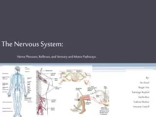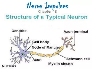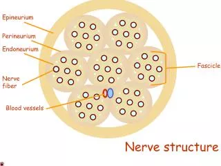
Nerve Plexuses
Nerve Plexuses. All ventral rami except T 2 -T 12 form interlacing nerve ___________________________called _ Plexuses are found in the cervical, brachial, lumbar, and sacral regions Each resulting branch of a plexus contains _. Nerve Plexuses.

Nerve Plexuses
E N D
Presentation Transcript
Nerve Plexuses • All ventral rami except T2-T12 form interlacing nerve ___________________________called _ • Plexuses are found in the cervical, brachial, lumbar, and sacral regions • Each resulting branch of a plexus contains _
Nerve Plexuses • Fibers travel to the periphery via several different routes • Each muscle receives a nerve supply _ • Damage to _____________________________________ cannot completely paralyze a muscle
Spinal Nerve Innervation: • The back is innervated by ______________________________ via several branches • The thorax is innervated by _________________________________ T1-T12as intercostal nerves • Intercostal nerves supply muscles of the ribs, anterolateral thorax, and abdominal wall
Cervical Plexus • The __________________________________ is formed by ventral rami of C1-C4 • Most branches are ________________________________ nerves of the neck, ear, back of head, and shoulders • The most important nerve of this plexus is the _ • The phrenic nerve is the major _
Brachial Plexus • Formed by C5-C8 and T1 (C4 and T2 may also contribute to this plexus) • It gives rise to the _
Brachial Plexus • There are four major branches of this plexus • _______________________________________ – five ventral rami (C5-T1) • _______________________________________– upper, middle, and lower, which form divisions • _______________________________________– anterior and posterior serve the front and back of the limb • _______________________________________– lateral, medial, and posterior fiber bundles
Brachial Plexus: Nerves • Axillary • Musculocutaneous • sends fibers to the biceps brachii and brachialis • branches to most of the flexor muscles of arm • supplies the flexor carpiulnaris and part of the flexor digitorumprofundus • Radial • innervates essentially all _
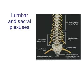
Lumbar Plexus • Arises from L1-L4 and innervates the _ • The major nerves are the _
Lumbar Plexus Figure 13.10
Sacral Plexus • Arises from L4-S4 and serves the buttock, lower limb, pelvic structures, and the perineum • The major nerve is the _ • The sciatic is actually composed of two nerves:
Innervation of Joints • Hilton’s law: any nerve serving a muscle that produces _____________________________ at a joint also innervates the _
Reflexes • A reflex is a _ • Reflexes may: • Be inborn _ • Involve only peripheral nerves and the _ • Involve higher brain centers as well
Reflex Arc • There are five components of a reflex arc • site of stimulus • transmits the afferent impulse to the CNS • either monosynaptic or polysynaptic region within the CNS • conducts efferent impulses from the integration center to an effector • muscle fiber or gland that responds to the efferent impulse
Reflex Arc Figure 13.14
Stretch and Deep Tendon Reflexes • For skeletal muscles to perform normally: • The Golgi tendon organs (_______________________________________) must constantly inform the brain as to the state of the muscle • Stretch reflexes initiated by muscle spindles must maintain healthy _
Muscle Spindles • Muscle spindles are wrapped with ______________________________________: primary sensory endings of type Ia fibers and secondary sensory endings of type II fibers • These regions are innervated by gamma () efferent fibers • Note: contractile muscle fibers are extrafusal fibers and are innervated by alpha () efferent fibers
Muscle Spindles Figure 13.15
Operation of the Muscle Spindles • __________________________________ the muscles activates the muscle spindle • There is an _________________________________________________________________________________ in Ia fibers • ___________________________________ the muscle ________________________________________on the muscle spindle • There is a decreased rate of action potential on Ia fibers
Operation of the Muscle Spindle Figure 13.17
Stretch Reflex • Stretching the muscle _ • Excited motor neurons of the spindle cause the stretched muscle to contract • Afferent impulses from the spindle result in inhibition of the antagonist • Example: • Tapping the patellar tendon _ • The quadriceps contract and the _
Golgi Tendon Reflex • The _____________________________ of the stretch reflex • ____________________________________ the muscle _ • Afferent Golgi tendon neurons are stimulated, neurons inhibit the contracting muscle, and the antagonistic muscle is activated • As a result, the contracting muscle relaxes and the antagonist contracts
Flexor and Crossed Extensor Reflexes • _____________________________________ is initiated by a _________________________ stimulus (actual or perceived) that causes automatic _____________________________________ of the threatened body part • The crossed extensor reflex has two parts • The stimulated side is _ • The _
Crossed Extensor Reflex Interneurons + + – + + – Efferent fibers Afferent fiber Efferent fibers Extensor inhibited Flexor inhibited Arm movements Flexes Flexor stimulated Extensor stimulated Extends Key: + Excitatory synapse – Inhibitory synapse Right arm (site of stimulus) Left arm (site of reciprocal activation)
Superficial Reflexes • Initiated by gentle ___________________________________ stimulation • Example: • __________________________________________ is initiated by stimulating the lateral aspect of the sole of the foot • The response is _ • Indirectly tests for proper ____________________________________________ functioning • _________________________________________ : abnormal plantar reflex indicating corticospinal damage where the great toe dorsiflexes and the smaller toes fan laterally
Autonomic Nervous System (ANS) • The ANS consists of motor neurons that: • Innervate _ • Make adjustments to ensure optimal support for body activities • Operate via _ • Have ____________________________________ as most of their _
ANS Versus Somatic Nervous System (SNS) • The ANS differs from the SNS in the following three areas • Efferent _ • Target organ responses
Effectors • The effectors of the _____________ are _ • The effectors of the ________________ are _
Efferent Pathways • _____________________________________ axons of the _______________________________________ extend from the CNS to the effector • Axons of the ANS are a _ • The ______________________________________ (first) neuron has a lightly myelinated axon • The _______________________________________ (second) neuron extends to an effector organ
Neurotransmitter Effects • All ____________________________________ neurons release _ • which has an _ • In the ANS: • Preganglionic fibers _ • Postganglionic fibers release • or _ • the effect is either _ • ANS effect depends on the • neurotransmitter released • and the _