Download
1 / 23
230 likes | 453 Vues
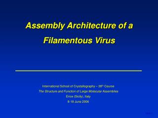
Assembly Architecture of a Filamentous Virus. International School of Crystallography – 38 th Course The Structure and Function of Large Molecular Assemblies Erice (Sicily), Italy 8-18 June 2006. 060611. Laboratory for Raman Spectroscopy of Biological Assemblies.

E N D
Assembly Architecture of a Filamentous Virus International School of Crystallography – 38th Course The Structure and Function of Large Molecular Assemblies Erice (Sicily), Italy 8-18 June 2006 060611
Laboratory for Raman Spectroscopy of Biological Assemblies UMKC School of Biological SciencesDivision of Cell Biology and Biophysics Current Recent James M. Benevides, Ph.D. Stacy A. Overman, Ph.D. Daniel Nemecek, Ph.D. Jessica Kawakami Cuong Tran Mitchell McGill John Schubler June Deng Ying Sun, Ph.D., University of Maryland Arantxa Rodriguez-Casado, Ph. D., CSIC, Spain Doinita Serban, Ph.D., Scripps Research Inst. Masamichi Tsuboi, Prof. Emeritus, U. Tokyo Collaborators Prof. Dennis H. Bamford & Dr. Roman Tuma, Biocenter, University of Helsinki Prof. Sherwood R. Casjens, Division of Molecular and Cellular Biosciences, University of Utah Prof. Edward H. Egelman, Department of Biochemistry and Molecular Genetics, University of Virginia Prof. Roger W. Hendrix, Department of Biological Sciences, University of Pittsburgh Prof. Jonathan A. King, Department of Biology, M.I.T. Prof. Peter E. Prevelige, Jr., Department of Microbiology, University of Alabama at Birmingham 060611
Outline of Presentation 1. Background filamentous bacterial viruses composition & morphology structural models: fiber x-ray diffraction solid state NMR spectroscopy 2. Emerging Structural Details electron cryomicroscopy Raman & polarized Raman future directions 060611
Morphogenesis of Filamentous Phage fd Life Cycle Elongation Process E. coli Courtesy of A. Kuhn Webster, R. E. (1996) 060611
Properties of Filamentous Viruses fd Pf1 Pf3 PH75 Symmetry class I (C5S2)a II (C1S5.4)b II (C1S5.4)b II (C1S5.4)c Length (nm) 880 1900 680 910 Ext. diam. (nm) 6.5 6.5 6.5 6.5 No. subunitsd 2750 7400 25002700 No. nucleotides 6408 7420 5800 6500 Nucl./subunit 2.4 1.0 2.4 2.4 Wt-% protein 87 94 86 87 a10 subunits per 32 Å helical repeat. Marvin et al. (2006) J. Mol. Biol. 355, 294-309. b27 subunits per 75 Å helical repeat. Welsh et al. (2000) Acta Cryst. D56, 137-150; Welsh et al. (1998) J. Mol. Biol. 283, 155-177. cPederson et al. (2001) J. Mol. Biol. 309, 401-421. dSequences: fd: AEGDDPAKAA FDSLQASATEYIGYAWAMVV VIVGATIGIK LFKKFTSKAS50 (5.24 kDa; pI = 6.3) Pf1: GVIDTSAVES AITDGQGDMK AIGGYIVGAL VILAVAGLIY SMLRKA46 (4.61 kDa; pI = 4.7) Pf3: MQSVITDVTG QLTAVQADIT TIGGAIIVLA AVVLGIRWIK AQFF44 (4.63 kDa; pI = 5.7) PH75: MDFNPSEVAS QVTNYIQAIA AAGVGVLALA IGLSAAWKYA KRFLKG46 (4.81 kDa; pI = 9.4) 060611
fd Architecture ssDNA core 65 Å 1/100th virion length Filament of 6.5 x 880 nm (PL = 2 μm) Coat of layered -helical subunits Arranged with 5-fold rotational symmetry Right-hand slew on capsid surface ssDNA packaged within (conformation?) Caspar & Makowski (1981) J. Mol. Biol. 145, 611-617. Day et al. (1988) Ann. Rev. Biophys. 17, 509-539. Marvin et al. (1994) J. Mol. Biol. 235, 260-286. fd (6.5 x 880 nm) 060611
Molecular Models of the fd Capsid solid state NMR fiber X-ray diffraction Zeri et al. (2003) PNAS 100, 6458-6463. Marvin et al. (2006) J. Mol. Biol. 355, 294-309. 060611
Summary Conflicting Models 1. Fiber diffraction (Marvin) - 1IFD(1994), 1IFI(1994), 1IFJ(1994),2C0W(2006) 2. Solid state NMR (Marvin) - 2C0X (2006) 3. Solid state NMR (Opella) - 1NH4 (2003) 4. Symmetry (Day) - unpublished Data-Based Structural Constraints Required 1. Orientation & registration of capsid subunits – cryoEM 2. Orientations of side chains – polarized Raman 3. Interactions of side chains – Raman 4. DNA conformation and base orientations – UVRR 060611
Outline of Presentation 1. Background filamentous bacterial viruses composition & morphology structural models: fiber x-ray diffraction solid state NMR spectroscopy 2. Emerging Structural Details electron cryomicroscopy Raman & polarized Raman future directions 060611
Electron Cryomicroscopy Approach Interactive Helical Real-Space Reconstruction (IHRSR) 060611
equilibrium persistence length: PL = 1.0 μm Electron Microscopy of fd Equilibrium persistence length (1.0 μm) is half of that reported previously. 060611
A 17.3 Å axial rise 34.6 ° twist angle 17.3 Å axial rise 34.6 ° twist angle B Electron Cryomicroscopy of WT fd ● Data collected from 84,310 segments of wild-type fd, each of 240 Å. ● Starting with a solid cylinder, IHRSR convergence to a single structure could not be achieved, regardless of initial symmetry. Thus, the data set was sub-divided. ● 25,440 segments converged to the same structure (A). ● 16,367 segments converged to a second structure (B). ● 42,503 segments failed to converge to either (A) or (B). Only structure B can be fitted with a continuously -helical subunit. 060611
Subunit density in B that is not apparent in A. Subunit -helix includes residues 6 - 48 Orientation of -helix in NMR and X-ray based models does not fit the capsid density of either A or B. Compatible with a continuously -helical subunit Density not fitted by a continuously -helical subunit NMR X-ray EM 90° Electron Cryomicroscopy of WT fd Structure A Structure B 060611
● Cryo-EM analysis has also been conducted on the fd mutant (Tyr 21 Met). Y21 Y24 W26 Electron Cryomicroscopy of Mutant fd ● For Y21M-fd, all selected segments (20,000) converged to either structure A (10,000) or structure B (10,000). Overman, et al., (1996) J. Mol. Biol. 259, 331-336; Tsuboi et al., (1996) Biochemistry 35, 10403-10410; Tsuboi et al., (2001) Biochemistry 40, 1238-1247. Structure A Structure B 060611
Cryo-EM Structure 060611
Cryo-EM Structure 060611
NMR X-ray EM Comparison with X-ray and NMR Models view from lumen view from exterior Structure B 060611
fd isotopomers (4) (2) (3) H X (1) (1) Donor only: I850/I830 = 0.30 (2) Donor & acceptor: I850/I830 = 1.25 (3) Acceptor only: I850/I830 = 2.50 (4) No H-bonding: I850/I830 = 6.70 830 850 Constraints from Raman Spectroscopy Solution Spectra: Assignments & Structural Interpretations Subunit: 90 ± 5% α-helical secondary structure Y21: no phenoxyl H-bonding (hydrophobic) Y24: no phenoxyl H-bonding (hydrophobic) W26: sequestered (hydrophobic); 2,1 (C-C-C-C1) = 108 ssDNA: C3-endo/anti deoxynucleoside conformations Overman et al., 1994, 1995, 1997, 1998, 1999, 2001, 2004 060611
fd polarized Raman spectra Icc Raman tensors + Ibb Constraints from Raman Spectroscopy Polarized Spectra on Oriented Specimens: 3-D Information Icc 4[sin2θ (r1 cos2 χ + r2 sin2 χ) + cos2 θ]2 –––– = –––––––––––––––––––––––––––––––––––––––––––––––– Ibb [cos2 θ (r1cos2 χ + r2 sin2 χ) + (r1 sin2 χ + r2 cos2 χ) + sin2 θ]2 Tsuboi & Thomas, 1996a, 1996b, 1997, 1998, 2001, 2005 060611
Y21 Y24 W26 Structural Constraints from Raman Data Polarized Spectra on Oriented Specimens: 3-D Information -Helix tilt angle: <h> = 16 4 ____________________________ Y21 orientation: = 71 5 = 36 5 (N-C-C-C) 1 = -68 (C-C-C-C) 2 = -71 ____________________________ Y24 orientation: = 71 5 = 36 5 (N-C-C-C) 1 = -47 (C-C-C-C) 2 = -31 ____________________________ W26 orientation: = 90 10 = 54 3 (N-C-C-C) 1 = -76 (C-C-C-C1) 2,1 = 108 4 Overman, et al., (1996) J. Mol. Biol. 259, 331-336; Tsuboi et al., (1996) Biochemistry 35, 10403-10410; Tsuboi et al., (2001) Biochemistry 40, 1238-1247. 060611
Summary and Prospects ● The first 3-D images of a native filamentous virus (fd) have been obtained. ● The fd capsid exhibits structural polymorphism (2 structures identified). ● A continuous α-helical subunit is consistent with only one of the structures. ● The fd capsid density is not well fitted by either X-ray (2C0W) or NMR (1NH4) models. ● IHRSR can address the pleiomorphic architecture of a filamentous virus and is expected to yield improved resolution of fd and class II (Pf1, Pf3, PH75) structures. 060611
NIH Grants GM50776 & GM54378 060611