Download
1 / 14
140 likes | 360 Vues
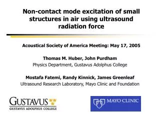
Non-contact mode excitation of small structures in air using ultrasound radiation force Acoustical Society of America Meeting: May 17, 2005. Thomas M. Huber, John Purdham Physics Department, Gustavus Adolphus College Mostafa Fatemi, Randy Kinnick, James Greenleaf

E N D
Non-contact mode excitation of small structures in air using ultrasound radiation forceAcoustical Society of America Meeting: May 17, 2005 Thomas M. Huber, John Purdham Physics Department, Gustavus Adolphus College Mostafa Fatemi, Randy Kinnick, James Greenleaf Ultrasound Research Laboratory, Mayo Clinic and Foundation
Introduction • Background material on ultrasound stimulated excitation • Study of different devices • MEMS mirror • Hard Drive HGA Suspension • MEMS Gyroscope • Conclusions
Ultrasound Stimulated Vibrometry • Pair of ultrasound beams directed at object • One ultrasound transducer differed from other by audio-range frequency • Difference frequency between ultrasound beams produces radiation force that causes vibration of object • Vibrations were detected using a Polytec laser Doppler vibrometer • In some experiments, comparison of ultrasound excitation and mechanical shaker
Experiment Details Confocal ultrasound transducer used • 600 kHz broadband (>100 kHz bandwidth) • 30 mm focal length; 1 mm focus spot size • Confocal (concentric elements with different frequencies) • Mounted on 3-D translation stage • Inner disk fixed at 500 kHz • Outer ring sweeps 501 – 520 kHz • Difference frequency of 1 kHz – 20 kHz Caused excitation of object
Device Tested: 2-d MEMS Mirror • Manufactured by Applied MEMS • Mirror is 3mm on Side - Gold plated Silicon • Three vibrational modes • X Axis torsion mode: 60 Hz • Z Axis torsion mode: 829 Hz • Transverse mode (forward/back): 329 Hz (incidental – not used for operation of mirror)
Selective Ultrasound Excitation of MEMS Mirror • Ultrasound focus ellipse about 1x1.5 mm • Focus position can be moved horizontally or vertically • Changing transducer position allows selective excitation • Upper figure: All modes present when focus near center of mirror. • Red line shows excitation using mechanical shaker. • Middle: X-torsional mode increases when ultrasound focus near top of mirror. • Bottom: Z-Torsional mode increases when focus near right edge
Selective Ultrasound Excitation of MEMS Mirror • X-Torsional mode peaks when focus near top/bottom of mirror • Transverse mode decreases as transducer moved vertically(smaller fraction of beam on mirror) • Ratio of amplitudes of X-Torsional to Transverse modes changes by over factor of 10x as vertical position is varied
Hard Drive HGA Suspension • HGA (Head Gimbal Assembly) suspension holds heads as they fly over the disk • Leading manufacturer: Hutchinson Technology, in Hutchinson, MN • Length about 5-10 mm, max. width about 2 mm, thickness of 25-100 μm • The suspensions are engineered to have specific vibrational modes. • Quality control involves measuring mode frequencies and deflection shapes(using mechanical shaker for excitation and vibrometer for measurement)
Ultrasound excitation of HGA Suspension • Goal: To determine whether vibrationalmodes of suspension can be excitedusing ultrasound radiation force • HGA Suspension was clampedand simply supported • Confocal ultrasound transducer usedto excite modes from 1 kHz to 50 kHz • Vibrometer measured resonance frequencies and deflection shapesat several ultrasound focus positions • Brüel & Kjær mechanical shakerused for comparison
Results for focused ultrasound excitation: HGA Suspension • Interference between ultrasound frequencies between 501 – 520 kHz and 500 kHz • Ultrasound focus (ellipse of about 1mm by 1.5 mm) centered on suspension (red curve) and towards edge of suspension (blue curve) Demonstrates feasibility of noncontact excitation using ultrasound radiation force. Selective Excitation: For ultrasound focus towards the edge (blue curve), large increase in amplitude of torsional modes at 6, 10, 13 and 15 kHz relative to the transverse modes at 2, 7, and 16 kHz.
Mode shapes determined using ultrasound excitation 2.0 kHz 7.2 kHz 6.0 kHz 10.8 kHz (Click on each image for animation of deflection shape)
Results for MEMS Gyroscope • Analog DevicesMEMS Gyroscope • Pair of Test Masses¾ mm squareseparated by 1.5 mm • 14 kHz resonance frequency Variation of Ultrasound Transducer Position Mechanical ShakerCan Produce Selective Excitation of Masses Shakes Entire Structure
Conclusions Ultrasound excitation shown to be feasible for modal analysis • Allows excitation of resonances from below 1 kHz to over 40 kHz • Parts such as MEMS mirror, gyroscope and HGA suspension • Completely non-contact for both excitation and measurement • Selective excitation of torsional/transverse modes • Selective excitation by moving ultrasound focus point • Similar selective excitation using phase shift between two transducers • Special thanks to • Hutchinson Technology • Applied MEMS • Analog Devices • Polytec Incorporated