External Ear Disease
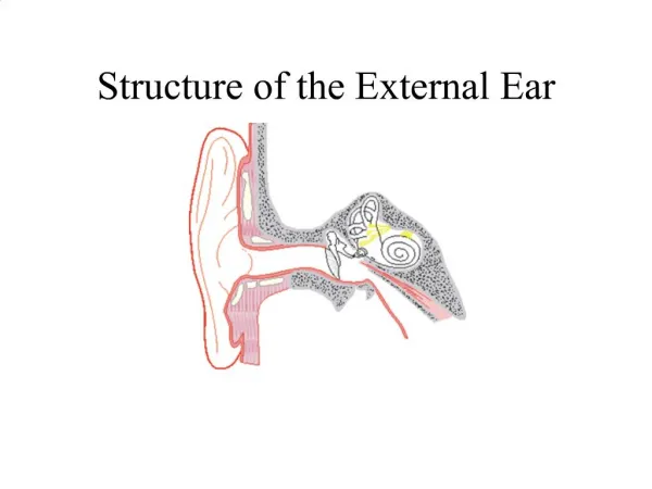
External Ear Disease . Dr. Lamia AlMaghrabi Consultant ENT King Saud Medical City . Anatomy . THE PINNA C omposed of cartilage with closely adherent perichondrium and skin. It is developed from six tubercles of the first branchial arch.


External Ear Disease
E N D
Presentation Transcript
External Ear Disease Dr. Lamia AlMaghrabi Consultant ENT King Saud Medical City
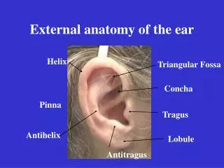
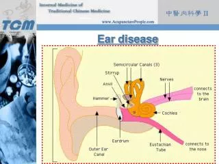
Anatomy • THE PINNA • Composed of cartilage with closely adherent perichondrium and skin. • It is developed from six tubercles of the first branchialarch. • Fistulae and accessory auricles result from failure of fusion of these tubercles.
THE EXTERNAL AUDITORY MEATUS • 25 mm in length. • Outer 1/3: • has a skeleton of cartilage, where it contains hairs and ceruminousglands. • Inner 2/3: • Bony skeleton, the skin of the inner part is exceedingly thin, adherent and sensitive. • At the medial end of the meatus there is the antero-inferior recess, in which wax, debris or foreign bodies may lodge.
THE TYMPANIC MEMBRANE • Composed of three layer: • skin, • fibrous tissue • mucosa. • The normal appearance of the membrane is pearly and opaque, with a well-defined light reflex due to its concave shape.
Congenital • Protruding ears • The underlying deformity is the absence of the antehelicalfold in the auricular cartilage. • Afflicted children are often teased mercilessly and surgical correction can be carried out after the age of four. • Operation consists of exposing the lateral aspect of the cartilage from behind the pinna and scoring it to produce a rounded fold
Accessory auricles • Accessory auricles are small tags, often containing cartilage, on a line between the angle of the mouth and the tragus. • They may be multiple.
Pre-auricular sinus • Pre-auricular sinus is a small blind pit that occurs commonly anterior to the root of the helix. • Sometimes bilateral. • Familial. • Recurrent infection • requires excision
Microtia • Failure of development of the pinna, may be associated with atresia of the ear canal. • Absence or severe malformation of the external ear, as in Treacher Collins syndrome, may be remedied by the fitting of prosthetic ears. • A bone-anchored hearing aid can be fitted at the same time, although it is often fitted at a much earlier age than prosthetic ears in order to allow speech development.
Congenital atresia: • may be of variable severity; • shallow blind pit or no cavity at all. • There may be associated absence of the pinna (microtia) and there may be absence or abnormality of the middle or inner ear. • In bilateral cases the cochlear function needs to be measured carefully. • If it is good, surgery may be considered. • In unilateral cases, it is of prime importance to assess the hearing in the unaffected ear. • If it is good, operation on the affected side is unnecessary.
TRAUMA Haematoma • Subperichondrialhaematoma of the pinna usually occurs as a result of a shearing blow. • The pinna is ballooned and the outline of the cartilage is lost. • If left untreated, severe deformity will result >>>cauliflower ear. • Treatment consists of evacuation of the clot and the reappositionof cartilage and perichondrium by pressure dressings or vacuumdrain.
AVULSION • Very rarely. • If the avulsed ear is preserved, reattachment may be possible.
INFLAMMATION • Acutedermatitis • Acute dermatitis of the pinna may occur as an extension of meatalinfection in otitis externa: • it is commonly caused by a sensitivity reaction to topically applied antibiotics, especially chloramphenicol or neomycin.
TREATMENT • 1 The ear canal should be adequately treated (q.v). • 2 If there is any suspicion of a sensitivity reaction, topical treatment with • antibiotics should be withdrawn. • 3 The ear may be treated with glycerine and ichthammol, or steroid ointment • may be applied sparingly. • 4 Severe cases may require admission to hospital.
Perichondritis • Perichondritismay follow injury to the cartilage and may be very destructive. • It may follow mastoid surgery or may follow ear piercing. • Treatment must be vigorous, with parenteral antibiotics and incision if necessary.
Chondrodermatitischronicishelicis • Chondrodermatitischronicishelicis occurs in the elderly as a painful ulcerated lesion on the rim of the helix. • It resembles a neoplasm and should be removed for histology.
OTITIS EXTERNA • Diffuse inflammation of the skin lining the external auditory meatus. • It may be bacterial or fungal (otomycosis). • Characterizedby: • irritation, desquamation, scanty discharge and tendency to relapse.
CAUSES: • Some people are particularly prone to otitis externa, often because of a narrow or tortuous external canal. • Swimming baths. • Poking the ear with a finger or towel further traumatizes the skin and introduces new organisms. • hotter climates than usual, where increased sweating is predisposing factor. • Underlying skin disease, such as eczema or psoriasis.
SYMPTOMS • 1 Irritation. • 2 Discharge (scanty). • 3 Pain (usually moderate, sometimes severe, increased by jaw movement). • 4 Deafness.
SIGNS: • 1 Meatal tenderness, especially on movement of the pinna or compression of the tragus. • 2 Moist debris, often smelly and keratotic, the removal of which reveals red desquamated skin and oedema of the meatal walls and often the tympanicmembrane.
MANAGEMENT • Scrupulous aural toilet is the key to successful treatment of otitis externa. • No medication will be effective if the ear is full of debris and pus. • Investigation • Investigation of the offending microorganism is essential. • A swab should be sent for culture and it is prudent to mention the possibility of fungal infection in your request, especially if the patient has already had topical antibiotic treatment.
Furunculosis • results from infection of a hair follicle and so must occur in the lateral part of the meatus. • The organism is usually Staphylococcus;
SYMPTOMS • Pain is as severe as that of renal colic and the patient may need pethidine. • The pain is made much worse by movement of the pinna or pressure on the tragus. • Deafness is usually slight and due to meatal occlusion by the furuncle.
Signs • There is often no visible lesion but the introduction of an aural speculum causes intense pain. • If the furuncle is larger, it will be seen as a red swelling in the outer meatus and there may be more than one furuncle present. • At a more advanced stage, the furuncle will be seen to be pointing or may present as a fluctuant abscess.
TREATMENT • The insertion of a wick soaked in 10% ichthammol in glycerine • Flucloxacillin should be given parenterally for 24 h, followed by oral medication. • Analgesics are necessary. • Recurrent cases are not common—exclude diabetes
TUMOURS • Squamous cell and basal cell carcinomas • These tumours occur usually on the upper edge of the pinna, and when small are easily treated by wedge excision. • Large tumours of the pinna or outer meatus will require more radical treatment, often with skin flaprepair.
Exostoses • small osteomata of the external auditory meatus. • Fairly common and usually bilateral. • They are much more common in those who swim a lot in cold water, although the reason is not known. • There may be 2 or 3 little tumours arising in each bony meatus. • Rate of growth is extremely slow and they may give rise to no symptoms, but if wax or debris accumulates between the tympanic membrane and the exostoses, In such cases, surgical removal of the exostoses may be indicated.