Download
1 / 39
390 likes | 749 Vues
Biology of Microorganisms. Presented by د. آصف احمد محمد جي مان فطاني بكاالوريوس الطب والجراحة (جامعة الملك عبدالعزيز) ماجستير الكائنات الدقيقة الطبية والجزيئية (جامعة مانشستر) دكتوراه الكائنات الدقيقة الطبية (جامعة مانشستر – بريطانيا) Dr Asif Jiman-Fatani, MB ChB, MSc, PhD (UK)

E N D
Biology of Microorganisms Presented by د. آصف احمد محمد جي مان فطاني بكاالوريوس الطب والجراحة (جامعة الملك عبدالعزيز) ماجستير الكائنات الدقيقة الطبية والجزيئية (جامعة مانشستر) دكتوراه الكائنات الدقيقة الطبية (جامعة مانشستر – بريطانيا) Dr Asif Jiman-Fatani, MB ChB, MSc, PhD (UK) Assistant Professor in Medical Microbiology, Faculty of Medicine, King Abdulaziz University Consultant Microbiologist Head, Clinical Microbiology Laboratories King Abdulaziz University Hospital 1431 H – 2010 G
Learning objectives • At the end of the lecture you should be able to: 1. Outline the main groups of microorganisms 2. Describe their important structural features 3. Know the medically significant microorganisms 4. Discuss the structural features that are important medically and for identification 5. Discuss how the metabolism and growth of microorganisms affect their infectivity and their control 6. Describe the indigenous flora of the human body, the areas colonized and the potential for infection
Naming Microorganisms For each organism 2 names (2 parts): Genusإسم الجنس- noun, always capitalized Species إسم النوع أو الفصيلة - adjective, lowercase Both italicized or underlined First letter may be used in an abbreviated version. Staphylococcus aureus (S. aureus) Bacillus anthracis (B. anthracis) Escherichia coli (E. coli) A common name is derived from historical use, e.g. pneumococcus for Streptococcus pneumoniae.
Major categories of microorganism • The main groups of microorganisms are: • Bacteria √ • Fungi √ • Helminths • Protozoa • Viruses √
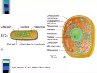
Single-celled organisms (prokaryotes أولية النواة ، بدائية النواة) Have a cell wall Contain both DNA and RNA Have no defined nucleus. May possess surface features such as pili (fimbriae), flagella or capsules. Do not have mitochondriaor other organelles (1). Bacteria
The Gram stain Many species can be defined as; Gram-positive, e.g. streptococci, or Gram-negative, e.g. Neisseria spp. Some organisms stain poorly with Gram stain but can be stained with other stains as mycobacteria (Ziehl-Neelsen stain). BacteriaStaining Reactions Gram Stain Gram Positive Gram Negative
Three shapes are seen: Spherical (coccus) مكورة Straight rod (bacillus) عصوية Curved or spiral ملتوية There is diversity within these groups; For example, cocci may be arranged in: Clusters (staphylococci), Chains (streptococci), or Pairs (pneumococci). BacteriaShape & Arrangement
BacteriaShapes (cont.) • Cocci المكورات • Gram-positive, e.g. staphylococci, streptococci • Gram-negative, e.g. Neisseria spp. • Bacilli العصويات • Gram-positive, e.g. clostridia - Bacillus spp., • Gram-negative, e.g. Escherichia coli - Pseudomonas spp. • Acid-fast, e.g. mycobacteria (Mycobacterium tuberculosis) • Spiral or curved rods e.g. vibrios, spirochaetes
Fungi possess DNA and RNA, a defined nucleus and a cell wall. There are two major types: Yeasts: Small, round, unicellular. Moulds: grow as filaments (hyphae) that may form mass (mycelium). Dimorphic fungi exist in both forms, e.g. Histoplasma. (2). Fungi الفطريات
Fungal reproduction • Asexual reproduction – spores are formed through budding or in conidia. • Sexual reproduction – spores are formed following fusion of male & female strains.
They grow inside a living cell (obligate intracellular parasites). Composed of a nucleic acid, either DNA or RNA, and a coat of protein subunits (capsomeres). A lipid envelope is found in some species. Viral particles have helical, icosahedral or no regular symmetry. (3). Viruses الفيروسات
Single-stranded DNA viruses, e.g. parvovirues Double-stranded DNA viruses, e.g. adenoviruses, herpesviruses, papovaviruses, poxviruses Single-stranded RNA viruses, e.g. bunyaviruses, coronaviruses, orthomyxoviruses, paramyxoviruses, picornaviruses, retroviruses, rhabdoviruses Double-stranded RNA viruses, e.g. neoviruses Segmented RNA viruses, e.g. arenaviruses Viruses (cont.)
Prokaryotic cell Eukaryotic cell
The bacterial cell is composed of the following structure Essential structure: • Cell wall. • Cytoplasmic membrane. • Intracytoplasmic structures : • Nuclear apparatus. • Ribosomes Non-essential structures: • Structures outside the cell wall • Capsules • Flagella • Fimbriae (pili). • Inclusion granules Other non-essentials: • Plasmids
Essential structure1. Bacterial cell wall Functions of the bacterial cell wall • Maintains the shape of bacteria. • Protects the cell from bursting in hypotonic solutions. • Protects the cell from mechanical disruption. • Provides a barrier against toxic chemical and biological agents. • Important in determining the cell's reaction to Gram stain. • Contains antigens that stimulate the patient’s antibody response. • Plays an essential role in cell division. • With the exception of mycoplasmas, all bacteria possess a cell wall
GRAM POSITIVE Lipoteichoic acid Peptidoglycan-teichoic acid Cytoplasmic membrane Cytoplasm GRAM NEGATIVE Lipopolysaccharide Porin Outer Membrane Braun lipoprotein Periplasmic space Inner (cytoplasmic) membrane Cytoplasm 18
Cell Wall The Gram-positive cell wall contains: Thick layer of peptidoglycan. Teichoic acids. The Gram-negative cell wall contains: Peptidoglycan is much thinner Lipoproteins. Outer membrane protein Lipopolysaccharides. Periplasmic space.
2. Cell Membrane The cell membrane is enclosed by the cell wall Mycoplasmas lack a cell wall and have an exposed cell membrane. Functions of the cytoplasmic membrane It plays a role in DNA replication. It is the site of respiration. It is a permeability barrier and contains proteins involved in selective and active transport of solutes. Active transport of ions (H+, Na+, K+, etc …) and nutrients into the cell.
3. Bacterial Chromosomal DNA • Single, supercoiled chromosome. • There is no nuclear membrane, no nucleolus, no mitotic apparatus, and no histones • The chromosome carries the genetic information to daughter cells and it is duplicated before cell division.
4. Ribosomes • Made of 60% ribosomal RNA & 40% protein • Consist of 2 subunits: large & small • Site of protein synthesis
1. Capsule External to the cell wall. Confers resistance to phagocytosis. 2. Pili (Fimbriae) Hair-like structures that protrude from the outer surface of some bacterial species Assist in adhesion to external surfaces. Non-essential structures
3. Flagella Flagella are long thin structuresthat protrude from the surface of some bacteria Organs of locomotion responsible for movement. 4. Inclusions granules Intracellular storage bodies. Examples: glycogen,, gas vesicles for floating, sulfur and polyphosphate granules
5. Plasmids • Extra-chromosomal DNA • Coding pathogenesis and antibiotic resistance factors
Spores الأبواغ Resting, dormant cells. Withstand extremes in heat, drying, freezing, radiation & chemicals not a means of reproduction Produced by some G+ genera. Have a 2-phase life cycle : Sporulation-formation of endospores. It contains calcium dipicolinate Germination- return to vegetative growth Pressurized steam at 120oC for 20-30 minutes will destroy.
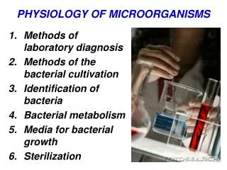
Bacterial Metabolism Factors that affect the rate of growth are: Temperature:Most bacterial species will grow at 37oC. 2. Hydrogen ion concentration (pH):Most pathogenic species can grow at pH 7.2 – 7.6. 3. Gaseous atmosphere: The gaseous environments used include: Aerobic: oxygen Anaerobic: lacks oxygen Microaerophilic: low oxygen Capnophilic: carbon dioxide.
Bacterial growth Bacterial growth follows recognisable stages. Lag phase: no increase in cell number Log phase: maximum increase in cell number Stationary phase: no net increase in cell number as a result of substrate limitation or inhibition by metabolite accumulation Death phase: decrease in cell number owing to toxic metabolites or substrate deprivation.
Air Outdoor aircontains bacteria, moulds and spores. Depend on the soil type, climate and population. Indoor aircontains organisms that are found in dust, droplets and droplet. Water Water acts as a vehicle for microorganisms that cause diseases, such as diarrhea, dysentery, enteric fever, cholera, hepatitis, etc Soil Soil exposure is important cases of tetanus, gas gangrene Bacteria are found in highest numbers in the layer penetrated by plant roots. Animals Some organisms are animal pathogens but can cause diseases in humans (Zoonotic disease)e.g. Brucella abortus (brucellosis in humans, septic abortion in domestic animals)
The indigenous human flora These organisms are normally found in harmless, close association with human body surfaces. The tissues, blood and internal body fluids of humans are normally sterile. Under certain circumstances, they can cause infection, e.g. Lowered host mechanisms e.g. immunosuppressed, diabetics, leukaemic patients. Alteration of the host tissues, e.g. Viridans streptococci may cause endocarditis after tooth extraction if the host has a predisposing heart lesion.
Skin Exposed areas are suitable for the growth of Staphylococcus epidermidis, coryneform bacilli, micrococci and low numbers of S. aureus. Numbers of bacteria are higher around hair shafts. Anaerobic bacteria (e.g. Propionibacterium acnes) are only found in anaerobic conditions of the sebaceous glands. An alteration in skin conditions that increases hydration or damages the surface (e.g. occlusion, high humidity, or chronic inflammatory conditions such as eczema and psoriasis) increases colonisation by organisms like Staphylococcus aureus.
Respiratory tract In the anterior nares, the species found are similar to those on the skin of the face. Staphylococcus aureus is present in up to 25-30% of adults. The nasopharynx contains streptococci, Non-pathogenic Neisseria spp., Streptococcus pneumoniae and Haemophilus influenzae Few microorganisms can be found below the larynx.
Gastrointestinal tract 1. Mouth Both α-haemolytic streptococci and non-pathogenic Neisseria are found on many surfaces. Streptococcus sanguis (important in the formation of dental caries) is present shortly after teeth eruption. Gingival crevice support the growth of Bacteroides spp.,fusiform bacteria and actinomycetes. 2. Stomach: Low pH and pepsin prevent the growth of most bacteria. 3. Small intestine:Motility keeps low numbers of organisms. 4. Large intestine: Anaerobic bacteria: Bacteroides fragilis Facultative bacteria: Escherichia coli and Enterococcus faecalis Other species present: staphylococci, clostridia, pseudomonads and yeasts.
Vagina In childhood, the organisms are aerobic bacteria such as Enterobacteriaceae, staphylococci and yeasts. At puberty (oestrogen) encourages the growth of lactobacilli;they create a low-pH Group B β-haemolyticstreptococci may be found colonising the adult vagina. At the menopause:Flora similar to that found before puberty, with an increase in Enterobacteriaceae.
Acquisition of the indigenous flora The baby’s colon is usually colonised within about 6-12 hours of birth. If the baby is breast-fed, this is mainly with bifidobacteria, and if bottle-fed, mainly with Enterobacteriaceae. Once an indigenous flora has been established, it is more difficult for new species to become established in the mouth or lower gastrointestinal tract. This has been called ‘colonisation resistance’.
Medical importance of the indigenous flora By definition, members of the indigenous human flora are not harmful in their normal habitat. However, under certain circumstances, they can cause infection, e.g. Colonic flora: urinary tract infection Skin flora: surgical wound infection Oral flora: dental caries, infective endocarditis
Medical importance of the indigenous flora (cont.) The alterations in indigenous flora seen when antibiotics are used can cause adverse effects in the patient such as: diarrhoea, colitis selection of antibiotic resistance secondary infection, e.g. candidiasis