Download
1 / 28
430 likes | 2.06k Vues
Erythrocyte metabolism. Alice Skoumalová. Erythrocytes deliver oxygen to body tissues and remove carbon dioxide and protons biconcave 7.7 μ m lack cell organelles 120 days women 4,2-5,4 million/ μ l , men 4,6-6,2 million/ μ l. The erythrocyte membrane

E N D
Erythrocyte metabolism Alice Skoumalová
Erythrocytes • deliver oxygen to body tissues and remove carbon dioxide and protons • biconcave 7.7μm • lack cell organelles • 120 days • women 4,2-5,4 million/μl, men 4,6-6,2 million/μl
The erythrocyte membrane 50% lipid bilayer (phospholipids, cholesterol) 50% proteins SDS-PAGE: separation of proteins (band 1-7) isolation and analysis (10 main proteins) Integral: Anion exchanger protein, Glycophorin A, B, C Peripheral: Spectrin, Ankyrin, Actin
Spectrin: the most prominent component (two isoforms α,β; a tetramer; a meshwork ) fixed to the membrane- ankyrin binding sites for several other proteins (glycophorin C, actin, band 4.1, adducin) This organization keeps the erythrocyte shape.
Haemoglobin 4 protein chains + 4 haem groups
Movements of the heme and the F helix during the T – R transition in hemoglobin:
Hemoglobin autooxidation • O2 binds Fe2+ - an intermediate structure - an electron is delocalized between the iron ion and the O2 • the side effect - every so often a molecule of oxyhaemoglobin undergoes decomposition and release superoxide Hem - Fe2+- O2 Hem - Fe3+ - O2•- • 3% of the haemoglobin undergoes oxidation every day Methemoglobin (Fe3+) is unable to bind O2 (methaemoglobin reductase)
Erythrocyte exceptions They lack organelles • no ATP production in oxidative phosphorylation • no ability to replace damaged lipids and proteins (low metabolic activities, with no ability to synthesize new proteins or lipids) Free radicals exposure • haemoglobin autoxidation (O2•- release) • a cell membrane rich in polyunsaturated fatty acids (susceptible to lipid peroxidation) • deformation in tiny capillaries; catalytic ions leakage (cause of lipid peroxidation)
Erythrocyte metabolism • Glucose as a source of energy • Glycolysis generates ATP and 2,3-bisphosphoglycerate • The pentose phosphate pathway produces NADPH • Glutathione synthesis- the antioxidant defence system
Glucose- source of energy Glucose transporter: • integral membrane protein (12 membrane-spanning helices) • a channel for the glucose transport • insulin-independent transporter Glycolysis in erythrocytes 1. Source of ATP • Lactate- the end product • Cover energy requirement 2. Generate 2,3-bisphosphoglycerate (2,3-BPG) • a major reaction pathway for the consumption of glucose in erythrocytes • the specific binding of 2,3-BPG to deoxyhemoglobin decreases the oxygen affinity of hemoglobin and facilites oxygen release in tissues
2,3-bisphosphoglycerate • Allosteric effector of haemoglobin: • binds to deoxyhaemoglobin (a central cavity capable of binding 2,3-BPG) • decreases haemoglobin‘s O2 affinity • Clinical aspects: • In people with high-altitude adaptation or smokers the concentration of 2,3-BPG in the blood is increased (low oxygen supply) • Fetal haemoglobin has low BPG affinity - the higher O2 affinity - facilitates the transfer of O2 to the fetus via the placenta
Glutathione Elimination of H2O2 and organic hydroperoxides 1. Cofactor for the glutathione peroxidase (removes H2O2 formed in erythrocytes) 2. Involved in ascorbic acid metabolism 3. Prevents protein –SH groups from oxidizing and cross-linking Glutathione peroxidase Gly Cys Glu Gly Cys Glu Gly Cys SH Glu + R-O-O-H S S + H2O + NADPH Glutathione reductase Reduced form of glutathione (monomer) Oxidized form of glutathione (dimer, disulphide)
The pentose phosphate pathway in erythrocytes • Generates NADPH - reduction of glutathione (eliminates H2O2 formed in erythrocytes) Clinical apect: • Glucose-6-phosphate dehydrogenase deficiency • Causes hemolytic anemia (decreased production of NADPH - reduced protection against oxidative stress - haemoglobin oxidation and Heinz bodies formation, membrane lipid peroxidation and hemolysis) • Hemolytic crises are evocated by drugs (primaquine, sulphonamide antibiotics) and foods (broad beans) • The most common enzyme deficiency disease in the world (100 million people)
Oxyhaemoglobin O2 Superoxide dismutase Haemoglobin Superoxide H2O2 Catalase Methaemoglobin reductase Methaemoglobin ½ O2+H2O Pentose phosphate pathway GSH NADP+ Glutathione reductase Glutathione peroxidase NADPH GSSG H2O GSH-reduced form; GSSG-oxidized form of glutathione
Haemoglobinopathy • abnormal structure of the haemoglobin (mutation) • large number of haemoglobin mutations, a fraction has deleterious effects • sickling, change in O2 affinity, heme loss or dissociation of tetramer • haemoglobin M and S, and thalassemias Haemoglobin M • replacement of the histidine (E8 or F7) in α or β-chain by the tyrosine • the iron in the heme group is in the Fe3+ state(methaemoglobin) stabilized by the tyrosine • methaemoglobin can not bind oxygen Thalassemias • genetic defects- α or β-chains are not produced (α or β-thalassemia)
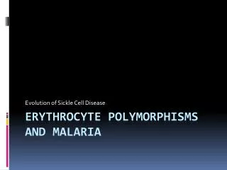
Haemoglobin S (sickle-cell) • Causes a sickle-cell anemia • Erythrocytes adopt an elongated sickle shape due to the aggregation of the haemoglobin S
Replacing Glu with the less polar amino acid Val - forming „an adhesive region“ of the β chain • Hg proteins aggregate into a long rodlike helical fiber Cross section
Red blood cells adopt a sickle shape in a consequence of the forming haemoglobin S fibers • The high incidence of sickle-cell disease coincides with a high incidence of malaria • Individuals heterozygous in haemoglobin S have a higher resistance to malaria; the malarial parasite spends a portion of its life cycle in red cells, and the increased fragility of the sickled cells tends to interrupt this cycle Scanning electron micrograph of a sickled erythrocyte. The haemoglobin S fibers can be seen within the distorted cell. The cell has ruptured and haemoglobin fibers are spilling out.
Glycosylated haemoglobin (HbA1) • formed by hemoglobin's exposure to high plasma levels of glucose • non-enzymatic glycolysation (glycation)- sugar bonding to a protein • normal level HbA1- 5%; a buildup of HbA1- increased glucose concentration • the HbA1 level is proportional to average blood glucose concentration over previous weeks; in individuals with poorly controlled diabetes, increases in the quantities of these glycated hemoglobins are noted (patients monitoring) Sugar CHO + NH2 CH2Protein Sugar CH N CH2 Protein Sugar CH2 NH CH2Protein Schiff base Amadori reaction Glycosylated protein
Summary • The erythrocyte membrane • Haemoglobin –O2 binding • Haemoglobin – allosteric efectors • Free radicals in erythrocytes • Metabolic pathways in erythrocytes • Haemoglobinopathy • Glycosylated haemoglobin
Pictures used in the presentation: Marks´ Basic Medical Biochemistry, A Clinical Approach, third edition, 2009 (M. Lieberman, A.D. Marks) Principles of Biochemistry, 2008, (Voet D, Voet J.G., and Pratt C.W) Color Atlas of Biochemistry, second edition, 2005 (J. Koolman and K.H. Roehm)