
The Digestive System
E N D
Presentation Transcript
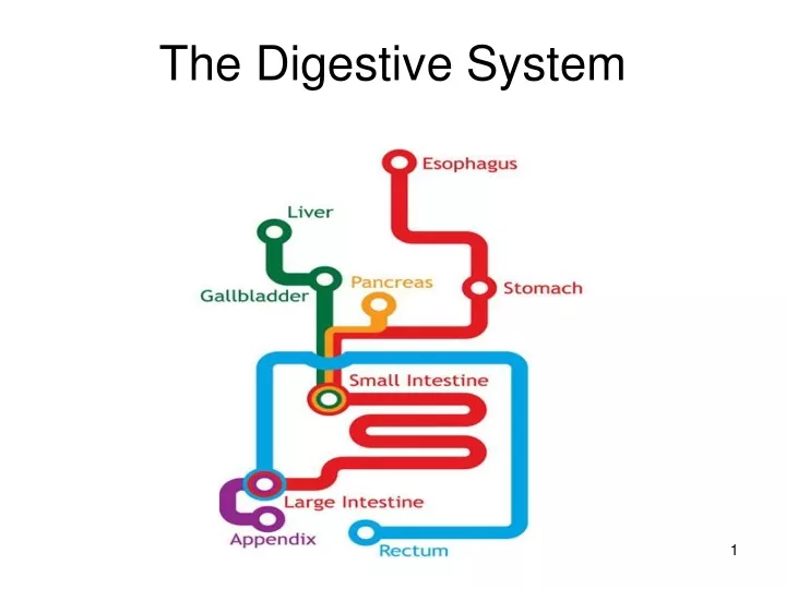
See fig. 12.1 p. 214; also table 12.1 p. 214 (overview of major digestion structures) Cardiac Sphincter Pyloric Sphincter
The Digestive Process Two key definitions: • Mechanical digestion is the physical breakdown of larger food ‘bits’ into smaller food ‘bits’. • Chemical digestion is the hydrolysis of macromolecules into smaller polymers or monomers. MOUTH • Both the Mechanical and Chemical Digestion of food begins in the mouth here, saliva is present. • The pH within the mouth cavity is 7 (neutral). a. Mechanical Digestion: -- food is chewed with TEETH (32 in total) in order to increase the surface area of the food so that chemical digestion is more efficient.
There are four types of teeth (see fig. 12.2 p. 215): i. Incisors: Scraping – 2 (per quarter of mouth) ii. Canines (Cuspids): Piercing – 1 iii. Premolars (Bicuspids): Ripping – 2 iv. Molars: Crushing – 3 (incl. “Wisdom” teeth) • Human Dental Formula = 2123 2123
The tongue manipulates the food to the different teeth-types and eventually into a ‘wet ball’ called a bolus. The tongue then pushes the bolus to the back of the mouth cavity to begin the process of swallowing. • The tongue also possesses taste buds which help to determine the quality of the food being consumed. Umami: savory, meaty, brothy.
b. Chemical Digestion: -- there are three types of salivary glands that are connected to the mouth cavity by ducts (‘connector tubes’). -- these glands SECRETE (produce and release) saliva into the mouth at the thought, smell, or taste of food. Salivary Ducts
Saliva consists of: i. 98% Water (pH of saliva is 7) ii. Mucus and electrolytes/ions iii. Various anti-microbial agents iv. Salivary Amylase (enzyme) Functions of Saliva: 1. To lubricate food making it easier to swallow and to protect the soft-tissue lining of the esophagus. 2. To inhibit the possible effects of pathogens in food. 3. To provide the digestive enzyme salivary amylase, which digests (hydrolyzes) STARCH into the disaccharide sugar MALTOSE. Salivary amylase functions optimally at a pH of 7. -- Not all of the starch that humans ingest is fully hydrolyzed in the mouth, some is swallowed along with the newly produced maltose.
Swallowing • Swallowing occurs in the pharynx (throat), a region between the mouth and the esophagus that receives food from the mouth and air from the nasal cavity. • Once the tongue pushes the bolus into the pharynx, swallowing is induced through a REFLEX action. • Two preventative measures also occur by reflex, simultaneous to the swallow-ing reflex, in order to disallow food from entering the air passages: Also: See Fig. 12.3 on p.216
As the bolus enters the pharynx, the soft palate (including the uvula) closes over the nasal passages at the roof of the pharynx. • At the same time, the epiglottis (flap of cartilage tissue) flops down over the glottis, which is the opening to the trachea, making the esophagus the primary receiver of the bolus. Thus, we do NOT breathe when we swallow. * The larynx is the first portion of the trachea and will be discussed in the Respiration Unit.
Esophagus (see fig. 12.4 p. 217) • The esophagus is a 25-30 cm muscular (smooth muscle) tube extending from the pharynx to the stomach; it is dorsal to the trachea (which is ventral). • “Dorsal” = Belly side; “Ventral” = Back/Spinal side. • No digestion occurs here, only the transport of the bolus to the stomach. • The inner lining of the esophagus (which lines the central cavity or lumen) is made up of mucosa cells which produce large amounts of mucus to constantly protect the esophagus from damage due to ‘coarse’, ‘sharp’, or acidic foods. • The bolus is squeezed rather tightly in the esophagus, so gravity is usually insufficient for transport (exception: liquids), but the mucus lining aids in easing the transport process.
The bolus then moves through the Cardiac Sphincter (‘Cardiac’ because it’s closer to the heart) which regulates a steady influx of food into the stomach. This sphincter acts as a VALVE that, for the most part, prevents the backflow of food into the esophagus. • Rhythmic smooth muscle contractions (known as PERISTALSIS) move the bolus down the esophagus toward the stomach. EXCEPTIONS: Vomiting (Reverse Peristalsis) and Heartburn. Read p. 217 “The Wall of the Digestive Tract” to learn a bit about tissue layers in the digestive tract.
Stomach • The stomach is a large, thick-walled, folded-walled, J-shaped, distensible organ that has the ability to expand to accommodate 2-4 L of food in its lumen (see fig. 12.5 p. 218). • It lies left of center beneath the diaphragm. Oblique Muscle Circular Muscle Longitudinal Muscle The term ‘gastric’ refers to the stomach.
Both Mechanical and Chemical Digestion occur within the lumen of the stomach: a. Mechanical Digestion i. Food is churned to break the bolus apart (thus increasing the Surface Area (SA) of the bolus) and to aid in its mixing with gastric juice. -- the churning occurs due to the contraction of the smooth muscle lining the stomach. -- there are three layers of smooth muscle: Circular, Longitudinal, and Oblique (diagonal). ii. Storage of Food (Mechanical Control): -- the stomach regulates the release of the ‘finished’ product into the small intestine.
b. Chemical Digestion -- the thought, smell, or taste of food triggers a nerve message from the medulla oblongata in the brain to various stomach cells. -- the result of this is the secretion of gastric juice (which contains ingredients from three different cells in the gastric pits of the stomach’s wall – more on these cells later). -- the actual presence of food (or saliva) in the stomach initiates the release of the hormone GASTRIN from the G cells of the stomach wall into the capillaries of the stomach. -- gastrin stimulates further release of gastric juice, in order to provide a sustained secretion that continues until food exits the stomach (essentially, gastrin takes over for the nerve message from the medulla oblongata) ** Nervous system STARTS the release… hormones SUSTAIN the release…
-- Gastric juice is composed of three key substances, each of which is secreted from a different stomach cell-type (thus, the medulla and gastrin have to signal all three cell types): • Mucus -- secreted by Mucosa (or Goblet) cells. -- mucus serves to line the interior wall of the stomach lumen in order to protect it from the caustic abilities of hydrochloric (HCl) acid and the protein-digesting abilities of the enzyme pepsin. -- the water portion of mucus helps to regulate temperature in the constantly active stomach since water has a high heat capacity.
Pepsinogen: -- a type of zymogen (a soon-to-be protein-digesting enzyme that is secreted in an inactive form). -- pepsinogen is a precursor to the important protein-digesting enzyme pepsin. -- pepsinogen is secreted by the Chief cells of the gastric pits into the stomach’s lumen. • Hydrochloric Acid (HCl) -- secreted by the Parietal cells of the gastric pits. -- has three major functions:
Disrupts the intercellular ‘glue’ (matrix) that binds cells together in meat and plant material (note: cell membranes do not touch, but they are ‘glued’ together with this matrix). HCl also denatures food proteins, thus ‘unfolding’ their tertiary structures. Both of these functions serve to increase the SA of food (thus, HCl contributes to mechanical digestion). • Lowers the pH of the stomach lumen to approx. 2-2.5 which kills most bacteria present in the ingested food. (Protection from disease). • Reacts with pepsinogen in order to activate it, forming the enzyme pepsin, which is an active PROTEASE. Pepsin then chemically digests food proteins down to smaller ‘proteins’ or polypeptides. However, since pepsin can only break peptide bonds adjacent to certain amino acids, protein hydrolysis is incomplete post-stomach.
GASTRIC PITS Stomach Lumen • The Mucosa, Chief, Parietal, and G cells are all located within stomach wall infoldings known as GASTRIC PITS (otherwise known as gastric glands). (secretes gastrin) It is important that the HCl and the pepsinogen are released from different cells because if they mixed prior to their release into the stomach lumen, pepsinogen would be activated into pepsin and the cells lining the stomach would be digested from the inside-out.
Pepsin’s optimal pH is 2-3 • The mucus protects the stomach wall from autodigestion by pepsin and denaturation by HCl. • Still, the stomach cells are constantly eroded, and mitosis must regenerate the cells every 3 days. • If there is a lack of mucus in a localized area (caused by stress (which can inhibit mucosa cell effectiveness) or a Helicobacter pylori bacterial infection (actually destroys mucus lining)), a gastric ulcer may form. • After stomach action, the remnants of swallowed food are now in liquid phase and are referred to as acid chyme (liquid). • The passage of acid chyme into the small intestine (duodenum) is regulated by the PYLORIC SPHINCTER. • It takes approx. 2-6 hours for a meal to completely exit the stomach.
A typical stomach ulcer There’s a hole in my stomach, dear Liza, dear Liza…
Small Intestine • A coiled, narrow tube about 6-7 meters in length and about 2-5 cm in diameter (called “small” because it’s narrow). • Divided into three sections: i. Duodenum: approx. 25-30 cm in length. -- lined with a thin layer of mucosa cells for mucus production. -- location of almost all chemical digestion. ii. Jejenum iii. Ileum Both the jejenum and ileum are primarily responsible for nutrient absorption, along with some ‘finishing touches’ to chemical digestion.
Most enzymatic (chemical) digestion of macromolecules occurs in the small intestine (SI) relative to the stomach and the mouth. • Furthermore, the absorption of the final products of digestion occurs in the SI with help from tiny ‘infoldings’ along its length called villi which increase the SA for absorption. Lumen of SI
Duodenum – Chemical Digestion • The duodenal walls possess chemoreceptors (chemical-sensitive nerve endings) that detect when as little as a drop of acid chyme enters the duodenum. • Once the chyme is detected, three hormones are released simultaneously from the cells lining the duodenum into the capillaries of the duodenum.
Hormone #1: Secretin - Travels through the blood to the pancreas (large gland that lies between the stomach and the SI). -Secretin signals the pancreas to release a Sodium bicarbonate (NaHCO3) solution through the pancreatic duct directly into the duodenum. - The NaHCO3 acts as a buffer to neutralize the acidic pH of the acid chyme in order to maintain a duodenal pH of about 8-10 (slightly basic in nature). - This is important for two reasons: i. The acid could eventually damage duodenal tissue if left un-neutralized (would erode relatively thin mucus layer relative to the stomach’s). ii. All enzymes that function in the duodenum and other regions of the SI require an optimal pH of 8-10.
Hormone #2: CCK (Cholecystokinin) -- travels through the blood to TWO locations: 1. Gall Bladder -- CCK signals the gall bladder to release bile into the bile duct taking it directly to the duodenum. -- bile is originally produced by the liver and is stored in the gall bladder. -- bile contains bile salts, which are important for the efficient digestion of fats. -- bile serves to EMULSIFY fats which means that large fat globules are physically broken down into smaller fat globules (‘spreads fats out’, so to speak). -- this increases the SA of the fats so that enzymes can collide (mix) with them more efficiently (more collisions) -- bile is NOT an enzyme!!! -- it contributes to Mechanical Digestion. -- bile also contains pigments that are byproducts of Red Blood Cell destruction in the liver (this allows these byproducts to exit the body in the feces and to not accumulate in the liver).
Pancreas -- CCK also signals the pancreas to release its digestive enzymes in the form of pancreatic juice. -- the juice is released into the pancreatic duct (with the NaHCO3) and into the duodenum to meet the incoming chyme (food). Emulsification of Fats (ignore the rest of the picture)
The Bile and Pancreatic Ducts Liver Pancreas ‘Direct’ connections to the Duodenum
Hormone #3: Enterogastrone • acid chyme in the duodenum also stimulates the duodenal cells to release the hormone enterogastrone (a steroid hormone). • enterogastrone travels to the stomach where it inhibits muscle contractions, thereby slowing down the entry of food into the duodenum. • this provides a suitable amount of time for the digestive processes to occur in the duodenum, thus ensuring that the process is as comprehensive as it needs to be.
Pancreas (An Accessory Organ) • The pancreas possesses two cell types: 1. Hormonal (Endocrine) Cells -- these cells produce the hormones insulin and glucagon that help to regulate blood-glucose levels (more on insulin later this unit). 2. Exocrine cells that secrete Pancreatic Juice -- pancreatic juice is released from the pancreas into the pancreatic duct directly into the duodenum. -- pancreatic juice contains the following substances which aid in chemical digestion of all four major macromolecules:
a. Trypsinogen (another zymogen): -- released into the duodenum and converted into the ACTIVE enzyme trypsin by a duodenal-produced enzyme enterokinase (duodenal equivalent of HCl in stomach, as it activates trypsinogen into trypsin) -- Trypsin digests the polypeptides (‘mini-proteins’) that resulted from stomach digestion into even smaller, more manageable, polypeptides. -- nevertheless, protein digestion is still incomplete. • Peptidases -- also released as zymogens and activated by the duodenum-released enterokinase. -- two types: i. Aminopeptidases ii. Carboxypeptidases
-- these enzymes digest the smaller polypeptides into individual amino acids by hydrolyzing them off of the chain one at a time. -- Seems onerous, but… -- increased SA of substrates (polypeptides) because of trypsin. -- aminopeptidase can cleave from amino end of chain while carboxypeptidase can cleave from the carboxyl end (meet at the middle). - even after this process, some dipeptides still exist!
Pancreatic Amylase -- an enzyme that digests the remaining starch to maltose (a disaccharide of two glucoses). -- does exactly the same job as Salivary Amylase except functions at a different pH. • Lipase -- an enzyme that digests the emulsified (high SA) fats (thanks to bile) into their fatty acid and glycerol constituents. • Nucleases -- various enzymes that digest DNA/RNA into individual nucleotides that we can use during DNA replication and RNA transcription. - however, some non-monomers of nucleic acids still exist even after nuclease action.
Once chemical digestion is completed, the jejenum and ileum portions of the small intestine must serve to ABSORB the monomers created by the digestive process. • However, two types of dimers still exist: • Maltose – a disaccharide made up of two glucose molecules. • Dipeptides – two amino acids joined together. *As well, some nucleic acids still require further digestion. • The digestion of these molecule-types does not occur within the duodenum.
Small Intestine (Jejenum/Ileum) • Topographically speaking, the lumen of the digestive tract is ‘outside’ the body (ie. It is a tunnel). • To ‘enter’ the body, the nutrients (amino acids, fatty acids, glycerol, nucleotides, glucose (eventually), vitamins, minerals, water) must cross the lining of the small intestine and enter the bloodstream. • Once in the blood, these nutrients can be delivered to all cells in the body, and not be eliminated as waste.
Food moves through the small intestine by peristalsis (SI is lined with smooth muscle). • The SI has a huge SA which maximizes the efficiency of absorption (600X more absorptive than that of a straight tube). • The jejenum/ileum are lined with villi (singular: villus) which are numerous infoldings/projections that increase the SA of the SI.
Furthermore, each villus possesses numerous microvilli which are further projections that serve to further increase the SA for absorption (the villi/microvilli both project towards the lumen of the SI). One villus
Each villus has its own capillary network as well as a lacteal (lymphatic capillary) (see fig 12.6 p. 219).
Villi Cells • The cells lining the villi are equipped with numerous mitochondria because active transport is often required for absorption. • The ‘lumen’ of the villi is the ECF between the villi cells and the capillaries/lacteals. • The villi cells also produce three enzymes and release them into their lumen just prior to absorption in order to complete digestion.
These three enzymes are: i. Maltase -- digests maltose to glucose within the villi lumen. ii. Dipeptidases -- digest dipeptides, producing amino acids. iii. Nucleosidases -- digest remaining small nucleic acid chains to individual nucleotides. ** all chemical digestion is now complete!!! • Once fatty acids and glycerol enter the villi lumen (ECF), they actually recombine to form fats which are too big to be absorbed by a capillary, but they are able to enter a lymphatic system capillary known as a lacteal.
So, fats initially enter the lymphatic system, but eventually enter into the circulatory system at the subclavian vein in the arm (where the lymphatic system forms a ‘Y’ with the circ. sys.). • What, therefore, do the blood capillaries within a villus absorb? • Amino acids • Glucose • Nucleotides • Vitamins • Minerals • Water
Large Intestine (Colon) • Once nutrients are absorbed, the remains consist of undigestible materials such as cellulose (fiber), excess water, and perhaps some nutrients that were consumed in excess. • These materials pass through the ileo-caecal valve into the large intestine (colon).
Anaerobic bacteria called E. coli live within the colon and feed on any unabsorbed nutrients that may exist there. • In return, E. coli produces various vitamins (B12, thiamine, riboflavin, K) that are normally deficient in the diet. E. coli • The major role of the colon is to reabsorb the water that was added to the food during the digestive process, thus solidifying the once liquid ‘stuff’. • The vitamins produced by E. coli are reabsorbed along with the water.
E. coli also begins the decomposition of the left-over food and converts it all to FECES with a changed colour, smell, and texture. • Read about diarrhea and constipation (p.221). • The rectum then stores the feces until defecation through the anus. ** the appendix is a small projection off of the colon which fights infections by accumulating toxins taken in through the mouth which somehow survived the stomach’s caustic lumen. * the appendix may become inflamed and may even burst.