( Failure to thrive( faltering growth
( Failure to thrive( faltering growth. Dr.Khalid Hama salih, Pediatrics specialist M.B.Ch.; D. C.H F.I.B.M.S.ped. ( FTT(faltering growth.

( Failure to thrive( faltering growth
E N D
Presentation Transcript
(Failure to thrive(faltering growth Dr.Khalid Hama salih, Pediatrics specialist M.B.Ch.; D. C.H F.I.B.M.S.ped
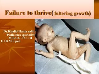
(FTT(faltering growth The term 'failure to thrive' is aclinical state of growth failure due to any causes,characterized by any one or more of the following features: (1) lack of growth i.e weight < 3red percentile (2) sustained weight loss( are less than the 80th percentile of median weight for height measurement) 3)Reduced growth velocity i.e weight dropping at least tow major percentile (e.g:from 75th to 25th ) Althuogh weight is the defining criteria for FTT,other growth parameter are freq\uently affected.
Differentiating the infant who is failing to thrive from a normal but small or thin baby is often a problem . Normal but short infants have no symptoms, are alert, responsive and happy, and their development is satisfactory. The parents may be short (low mid-parental height) or the infant may have been extremely preterm or growth-restricted at birth. Any intercurrent illness will be accompanied by a temporary failure to gain weight
epidemiology International International problems of poverty and hunger occur in many nations. No sex predilection is important to note No racial predilection is noted Age children from infancy through the toddler period.
Etiology: Non organic FTT: 80% due to emotional or nutritional deprivation Organic FTT: a).Decreased food intake,dispite availabilityeg:mechanicalcause, neuromusculardisease b)Impaired digestion/absorption c)Increase metabolic requirement e.g:chronic infection d)Increase losses of ingestedfood e.g:chronic diarrhea,vomiting
Non-organic : • a)Emotional deprivion • maternal death /chronic illness/separation • family disharmony • unwanted child • b)nutritional:protein-energy malnutrition • Organic: • infection:TB,chronicmalaria,kala azar ,AIDS • GIT : cleft palate, GERD,malabsorption • Cystic fibrosis • Hepatic: chronic liver • respiratory: recurrent infection, asthma, chronic lung disease • CVS: HF,CHD, infective endocarditis • CNS:CP, • Renal: chronic RTA,UTI • Endocrine: GH deficiency, CAH • Hematological:severe anemia,malignancy
Clinical manifestation: 1.Malnutrition e.g growthfailure,anemia,vitamin&mineral deficiency 2.Behavioral change:apathy,social withdrawal,poor eye contact 3.Developmental retardation 4.Recurrent &persistant infction 5.Sign of primary disease
management 1.Nutritional treatment is based on aggressive feeding to prevent cognitive loss. Most children require 100-120 kcal/kg/day, but this may be increased to achieve catch-up dietary instructions should include the following: • Eliminate empty calories from items such as soda or other high sugar drinks. • Schedule regular meals and snacks (usually 3 meals and 2 snacks per day). No grazing between meals. • Offer solids before liquids. • Consider fortifying calories with extra oils and carbohydrates. • Increase protein. • Consider vitamin and/or mineral supplements, especially A and iron.. • Avoid distractions, such as television,at meal time.
2 Observation of feeding is very important. Pay careful attention to the following: Maternal (caregiver) attachment during the feeding process; evaluation of signs of maternal attachment (eye contact, vocalizations) Evaluation of the child-parent dyad (eg, conflict over eating related to poor limit setting, or meal time disruption) The perception of parents and/or caregivers regarding the problem Feeding techniques (forced feeding) A 72-hour diet diary that includes the following can be helpful: Details relative to growth from breastfeeding or bottle-feeding Formula preparation and amounts provided Time and amount of feedings (eg, 5 oz of Enfamil) Behaviors of infant or child during feeding or nursing
Failure to thrive Initial evaluation(blood ,urine stool,CXR) Trial feedind (for 2 week) Wt gain satisfactory NonorganicFTT Unsatisfactory Organic FTT poor intake(abnormal suck,chew,sw Good intake chronic infection,systemic disease
Background Rickets is a disease of growing bone that is unique to children and adolescents. Rickets signifies a failure in mineralisation of the growing bone or osteoid tissue. Failure of mature bone to mineralise is osteomalacia.
Vitamin D deficiency rickets occurs when the metabolites of vitamin D are deficient. Less commonly, a dietary deficiency of calcium or phosphorus may also produce rickets
Vitamin D-3 (cholecalciferol) is formed in the skin from a derivative of cholesterol under the stimulus of ultraviolet-B light. Natural nutritional sources of vitamin D are limited primarily to fatty, fish liver oil.
Source of vitamin D Ultraviolet light fish liver oil Ergosterol (vitamin D-2) Dairy milk is fortified with vitamin D (400 IU/L) Human milk contains little vitamin D(less than 20-40 IU/L)
Cholecalciferol (i.e., vitamin D-3) is formed in the skin from 7-dehydrocholesterol.
This steroid undergoes hydroxylation in 2 steps. Occurs at position 25 in the liver, producing calcidiol (25-hydroxycholecalciferol) The first hydroxylation
The second hydroxylation Occurs in the kidney at the 1 position, where it undergoes hydroxylation to the active metabolite calcitriol (1,25-dihydroxycholecalciferol )
Calcitriol Acts at 3 known sites to tightly regulate calcium metabolism: (1)it promotes absorption of calcium and phosphorus from the intestine (2) it increases reabsorption of phosphate in the kidney (3) it acts on bone to release calcium and phosphate. Calcitriol may also directly facilitate calcification. These actions result in an increase in the concentrations of calcium and phosphorus in extracellular fluid.
Epidemiology The frequency increasing internationally 1.Children to wear sunscreen while outdoors 2.Children spend more time indoors watching television or playing electronic games, instead of playing outdoors
Aetiology Causes of rickets Nutritional (primary); • Dark-skinned people • Decreased exposure to sunlight, • Maternal vitamin D deficiency • Diets low in calcium, phosphorus and vitamin D, e.g. exclusive breast-feeding into late infancy • Macrobiotic, strict vegan diets Intestinal malabsorption: • Defective production of 25(OH)D3 - liver disease • Increased metabolism of 25(OH)D3 - enzyme induction by anticonvulsants Defective production of 1,25(OH)2D3 Hereditary type I vitamin D-resistant (or dependent) rickets (mutation which abolishes activity of renal hydroxylase) • Familial (X-linked) hypophosphataemic rickets (renal tubular defect in phosphate transport) • Chronic renal disease • Fanconi syndrome (renal loss of phosphate) Target organ resistance to 1,25(OH)2D3 Hereditary vitamin D-dependent rickets type II (due to mutations in vitamin D receptor gene)
Clinical features of rickets : • Misery • Failure to thrive/short stature • Frontal bossing of skull • Craniotabes • Delayed closure of anterior fontanelle • Delayed dentition • Rickety rosary • Harrison's sulcus • Expansion of metaphyses (especially wrist) • Bowing of weight-bearing bones • Hypotonia • Seizures (late
Knock knee deformity (genu valgum) Bowleg deformity (genu varum) Wrist enlargement Frontal bossing Rib beading (rachitic rosary) Harrison's sulcus and pot belly Tibial bowing
Differential Diagnoses • Hypophosphatasia • Hypophosphatemic vitamin D–resistant rickets. • Severe calcium deficiency • Severe phosphorus deficiency
Approach Considerations Serum measurements in the workup for rickets may include the following: 1.Calcium.2.Phosphorus.3.Alkaline phosphatase4.Parathyroidhormone 5.25-hydroxy vitamin D 6.1,25-dihydroxyvitamin D Radiography is indicated in patients with rickets
Serum Chemistry Calcium (ionized fraction) is low Calcidiol (25-hydroxy vitamin D) is low Parathyroid hormone is elevated Phosphorus level is invariably low for age Alkaline phosphatase levels are uniformly elevated.
Radiography • Cupping of the metaphysis • Fraying of the edge • Widening of the osteoid tissue • Hypominiralization of bones
Treatment & Management • Nutritional rickets is managed by advice about a balanced diet, correction of predisposing risk factors and by the daily administration of vitamin D3.
Treatment & Management Treatment for rickets may be administered gradually over several months or in a single-day dose of 15,000 mcg (600,000 U) of vitamin D • If the gradual method is chosen, 125-250 mcg (5000-10,000 U) is given daily for 2-3 months until: • Healing is well established • increasing vitamin D levels • Alkaline phosphatase concentration is approaching the reference range(, but complete reversal of bony deformities may take years)