Introductory Questions #1
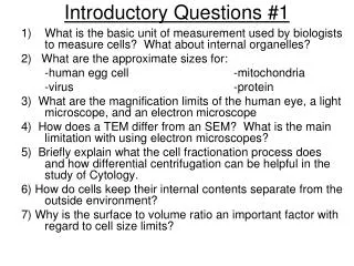
Introductory Questions #1. 1) What is the basic unit of measurement used by biologists to measure cells? What about internal organelles? 2) What are the approximate sizes for: -human egg cell -mitochondria -virus -protein

Introductory Questions #1
E N D
Presentation Transcript
Introductory Questions #1 1) What is the basic unit of measurement used by biologists to measure cells? What about internal organelles? 2) What are the approximate sizes for: -human egg cell -mitochondria -virus -protein 3) What are the magnification limits of the human eye, a light microscope, and an electron microscope 4) How does a TEM differ from an SEM? What is the main limitation with using electron microscopes? 5) Briefly explain what the cell fractionation process does and how differential centrifugation can be helpful in the study of Cytology. 6) How do cells keep their internal contents separate from the outside environment? 7) Why is the surface to volume ratio an important factor with regard to cell size limits?
Introductory Question #2 1) Name three structures found in prokaryotic cells, eukaryotic plant cells, and eukaryotic animal cells. 2) Name the three layers that surround and protect a prokaryotic cell. Why are prokaryotes considered to be “simple” cells and eukaryotic are called “complex” cells? Matching Ex. Cellular respiration A. Nucleolus Digests waste, worn out organelles B. Endoplasmic Ret. Produces rRNA and ribosomes C. Ribosomes Produces H2O2 D. Golgi Complex Forms Mitotic spindle in Mitosis E. Lysosmes Site for protein synthesis F. Peroxisomes Site for the synthesis of lipids G. Mitochondria Modifies, packages and ships protein H. Centrioles
IQ #3 What purpose do vesicles serve in the cell? • Name all of the organelles that are a part of the endomembrane system. 4) Explain how the rough ER is different from the smooth ER, 5) How is a lysosome different from a peroxisome? 6) What do the chaperone proteins in the ER do?
Introductory Questions # 4 • Name the people that helped to develop the cell theory. What contribution did each person make (what did they discover)?
Thank you for your response. Be sure the students are registering only once. We have had reports of students registering each time they need to access the Companion Website.Follow the registration steps below to access your textbook resources. Each teacher code will work for 2 teachers and each student code will work for up to 50 students. 1. You MUST begin the registration at - http://www.phschool.com/access/2. Click on "Covered Titles," then click on your title from the list3. Choose Teacher or Student Registration4. Click I Accept at the bottom of the License Agreement page 5. Access Information - * Enter or Create your username & password * Enter the appropriate access code below: (5th) SSNAST-PRINK-ASPIC-VARNA-GABBY-RITES(6th) SSNAST-TTBBO-ASPIC-VARNA-RIGOT-SITESSSNAST-MUSIL-ASPIC-VARNA-BEGUN-EANESSSNAST-BLIDA-ASPIC-VARNA-GIBBY-SIRESSSNAST-SHALL-ASPIC-VARNA-SOTTO-VOTES6. Account Information - complete or verify your name & school information7. Confirmation & Summary - you will receive a confirmation email which contains a link to the Companion Website.
A Tour of the Cell Chapter 6 (Pgs 94-123) • History & discoveries • Microscopy • Limits to Cell Size (Surface area to volume ratio) • Cell Fractionation (Structure & Function of Organelles) • Prokaryotic vs.Eukaryotic • Plant cells vs. Animal • Endomembrane System • Cytoskeleton • Intercellular junctions
History & Discovery of Cells • Anton Van Leeuwenhoek (pond water 1600’s) • Robert Hooke (Cork Cells, 1665) • Robert Brown (Nucleus, 1833) • Matthias Schleiden (Plant Cells, 1838) • Theodor Schwann (Animal Cells, 1839) • Rudolf Virchow (All Cells arise from other cells) • Cell Theory: 3 aspects
Below is a list of the most common units of length biologists use (metric) Table 4.2
Biological Size and Cell Diversity (Pg. 95) Human Eye: 1mm - meter+ LM: 1m – 1mm EM: 1nm – 1mm Chicken Egg (largest cell) Mitochondria (1m) Ribosomes (20-30 nm) Viruses (80-100 nm)
Microscopes provide windows to the world of the cell • The light microscope enables us to see the overall shape and structure of a cell Image seen by viewer Eyepiece Ocularlens Objective lens Specimen Condenser lens Light source Figure 4.1A
Scanning electron microscope (SEM) • Scanning electron micrograph of cilia Figure 4.1B
Transmission electron microscope (TEM) • Transmission electron micrograph of cilia Figure 4.1C
Cytology: science/study of cells • Light microscopy • resolving power~ measure of clarity • Electron microscopy (2 types) •TEM~ electron beam to study cell ultrastructure •SEM~ electron beam to study cell surfaces • Cell fractionation~ cell separation; organelle study • Ultracentrifuges~ cell fractionation; 130,000 rpm
Cell Fractionation • Physically separates and purifies cell parts • Spun in a centrifuge (up to 500,000 rpm) • Two fractions: supernatant & pellet • Differential: successively at higher speeds • Density gradient: forms bands in tube according to density differences of organelles
Cell Size • Is it more advantageous to be a single cell that is large or to be broken down into several small cells ? (Explain your answer)
A small cell has a greater ratio of surface area to volume than a large cell of the same shape 30 µm 10 µm Surface areaof one large cube= 5,400 µm2 Total surface areaof 27 small cubes= 16,200 µm2 Figure 4.3
Cell size - (surface area:volume) • As cell size increases, the surface area to volume ratio decreases (sa/vol) • Rates of chemical exchange may then be inadequate for cell size • Cell size, therefore, remains small
Natural laws limit cell size • At minimum, a cell must be large enough to house the parts it needs to survive and reproduce • The maximum size of a cell is limited by the amount of surface needed to obtain nutrients from the environment and dispose of wastes
The Prokaryotic Cell-(See Fig. pg 98)(Also See Pages 534-547 in Ch. 27) • Characteristics include: • No true distinct nucleus • Have a “Nucleoid” region = DNA & Plasmids • No complex, membranous organelles (Ribosomes only) • Most have rigid cell walls • Flagella (rotary type structure & not composed w/microtubules) • Some have pigments (autotrophic) • Classified according to their metabolic needs • Eubacteria & Archeabacteria • Some have sticky capsules, pili, peptidoglycan, Endospores • Asexually Reproduce: Binary Fission, Budding, Fragmentation • Genetic Material Can be exchanged by 3 mechanisms: • Transformation, Transduction, and Conjugation
The Eukaryotic Cell • “Eu” = true “Karyo” = kernal (nucleus) • Protists, Plants, Fungi, and Animals • Internal Membrane System • Has many membranous organelles (Table 4.1) that include: -Nucleus -Lysosomes -Golgi complex -Endoplasmic reticulum (R & S) -Mitochondria -Chloroplast (plastids) -Peroxisomes (glyoxysomes) -Vesicles -Vacuole (food, contractile)-Ribosomes • Cytoskeleton: microtubules, microfilaments, and int. filaments • Centrioles (nine triplets of microtubules) • Cilia & Flagella (9+2 microtubule arrangement) • Extracellular matrix (ECM)-proteins & carbodydrate -glycoproteins -glycolipids -integrins -fibronectins -collagen
Roughendoplasmicreticulum Nucleus Ribosomes Smoothendoplasmicreticulum Golgiapparatus Microtubule Centralvacuole Not inanimalcells Intermediatefilament Cytoskeleton Chloroplast Microfilament Cell wall Mitochondrion Peroxisome Plasma membrane Figure 4.5B
Smooth endoplasmicreticulum Nucleus Roughendoplasmicreticulum • An animal cell Flagellum (exception is some plants) Not in most plant cells Lysosome Centriole Ribosomes Peroxisome Golgiapparatus Microtubule Plasma membrane Cytoskeleton Intermediatefilament Microfilament Mitochondrion Figure 4.5A
Endomembrane Function http://users.uma.maine.edu/SusanBaker/nucleus_endo.html
Nucleus, Ribosomes, Rough & Smooth ER, Flow of Genetic information and protein Synthesis
Nucleus (Pg. 103) Control Center of the Cell Genetic material: •chromatin •chromosomes Nucleolus: ribosome synthesis Double membrane envelope with pores 1st part of Protein synthesis: Transcription (DNAmRNA) Nuclear pores
NUCLEUS Chromatin Two membranesof nuclearenvelope Nucleolus Pore ROUGHENDOPLASMICRETICULUM Ribosomes Figure 4.6
Ribosomes • Manufactures Protein • Free •cytosol; •protein function in cell • Bound •endoplasmic reticulum; •membranes, organelles, and export
Endoplasmic Reticulum (pg. 105) Endoplasmic reticulum (ER) • Continuous with nuclear envelope Smooth ER •no ribosomes; •synthesis of lipids •metabolism of carbohydrates • detoxification of drugs and poisons Rough ER •with ribosomes •synthesis of secretory proteins (glycoproteins), membrane production **Found extensively in Pancreas
Transport vesiclebuds off 4 Ribosome Secretory(glyco-) proteininside transportvesicle Sugarchain 3 Glycoprotein 1 2 ROUGH ER Polypeptide Rough Endoplasmic Reticulum makes membrane and proteins • The rough ER manufactures membranes • Ribosomes on its surface produce proteins Figure 4.8
SMOOTH ER ROUGHER Nuclearenvelope Ribosomes SMOOTH ER ROUGH ER Figure 4.9
Golgi Complex (pg. 106) • Golgi apparatus • •ER products are modified, stored, and then shipped • Cisternae: flattened membranous sacs • trans face (shipping) & cis face (receiving) • Transport vesicles
The Golgi apparatus finishes, sorts, and ships cell products • The Golgi apparatus consists of stacks of membranous sacs • These receive and modify ER products, then send them on to other organelles or to the cell membrane
The Golgi apparatus Golgi apparatus Golgiapparatus “Receiving” side ofGolgi apparatus Transportvesiclefrom ER Newvesicleforming “Shipping”side of Golgiapparatus Transport vesiclefrom the Golgi Figure 4.10
Lysosomes digest the cell’s food and wastes (Pg.107) • Lysosomes are sacs of digestive enzymes budded off the Golgi LYSOSOME Nucleus Figure 4.11A
Contain lysosomal enzymes (hydrolytic enzymes) • digests food molecules (macromolecules) • destroys bacteria • recycles damaged organelles • function in embryonic development in animals • undergoes phagocytosis & engulfs material • Recycle cell’s own organic material • **Found extensively in Macrophages (WBC’s) Lysosomes:
Rough ER Transport vesicle(containing inactivehydrolytic enzymes) Plasmamembrane Golgiapparatus Engulfmentof particle Lysosomeengulfingdamagedorganelle “Food” LYSOSOMES Digestion Foodvacuole Figure 4.11B
Lysosomes can cause Fatal Diseases • Lysosomal Storage Diseases are hereditary that interfere with other cellular functions *Examples: Pompe’s disease (build up of glycogen) Tay-Sachs disease (lipid build up) (Pgs. 93, 331)
Vacuoles -Membrane-bound sacs (larger than vesicles) -Food (phagocytosis) -Contractile (pump excess water) -Central (storage in plants) -Tonoplast membrane
Vacuoles function in the general maintenance of the cell • Plant cells contain a large central vacuole • The vacuole has lysosomal and storage functions Centralvacuole Nucleus Figure 4.13A
Peroxisomes (Pg. 111) • Single membrane • Oxidative organelle ***strips e-’s (H’s) from substances • Produce hydrogen peroxide (H2O2) in cells • Metabolism of fatty acids; detoxification of alcohol (liver) • Hydrogen peroxide then converted to water
Mitochondria & Chloroplasts -Energy Harvesting Organelles
Mitochondria harvest chemical Energy from food • Site for Cellular Respiration---Prod. of ATP • Uses O2 to extract energy from sugar, fats, and other molecules • Found in cells that are motile and contractible • Has a double membrane • Has Convoluted inner membranes: Cristae • Two spaces: Matrix & intermembrane space • Not part of the endomembrane system • Has its own DNA and rbosomes (able to regenerate & divide)---Semiautonomous
MITOCHONDRION Outermembrane Intermembranespace Innermembrane Cristae Matrix Figure 4.16
Chloroplasts convert solar energy to chemical energy • Chloroplasts are found in plants and some protists • Chloroplasts convert solar energy to chemical energy in sugars Chloroplast Stroma Inner and outer membranes Granum Intermembranespace Figure 4.15
The Chloroplast(pg. 111) • Site for Photosysnthesis: combines CO2 & H2O • Converts solar energy into chemical energy (sugar molecules) • A Type of Plastid • Three types: (Amyloplastid, chromoplast, and chloroplast) • Double membrane w/ thylakoids (flattened disks) • Grana (stacked thylakoids) • Three compartments -Stroma -Intermembrane space -Within the thylakoid membranes • Has its own DNA
The Cytoskeleton (pg. 112-113) -Fibrous proteins (actin & tubulin) -Support, cell motility, biochemical regulation, organelle movement -Microtubules: •thickest ( nm) •tubulin protein; •shape, support, transport, chromosome separation -Microfilaments: •thinnest ( nm) •actin protein filaments; •motility, cell division, shape -Intermediate filaments: • middle diameter; •keratin; •shape, nucleus anchorage