Hearing Disorders
Murad Al-momani, Ph.D., CCC-A, FAAA American Board of Audiology. Hearing Disorders . Hearing loss. Hearing loss is defined as having one or more frequencies out of the normal hearing range and it has degrees.

Hearing Disorders
E N D
Presentation Transcript
Murad Al-momani, Ph.D., CCC-A, FAAA American Board of Audiology Hearing Disorders
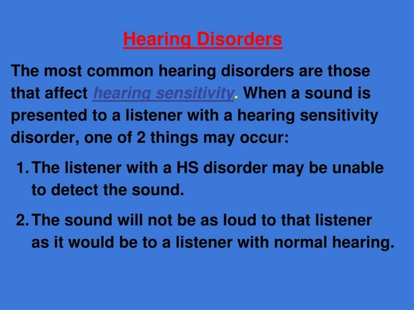
Hearing loss • Hearing loss is defined as having one or more frequencies out of the normal hearing range and it has degrees. • The more sever the hearing loss is, the more effect will be on the overall functioning of the individual with hearing loss. • But, even slight hearing loss can impede the development and acquisition of the normal language.
Types of Hearing loss • Conductive hearing loss: a failure in the efficient conduction of sound waves through the outer ear, tympanic membrane (eardrum) or middle ears (ossicles).
Causes of conductive hearing loss • Earwax, also known by the medical term cerumen, is a yellowish, waxy substance secreted in the ear canal of humans and many other mammals. • It plays an important role in the human ear canal, assisting in cleaning and lubrication, and also provides some protection from bacteria, fungi, and insects. • Excess or impacted cerumen can press against the eardrum and/or occlude the external auditory canal and impair hearing
Otitis media is an inflammation of the middle ear: the space behind the ear drum. Otitis media is very common in childhood, and includes acute and chronic conditions; all of which involve inflammation of the ear drum (tympanic membrane), and are usually associated with a buildup of fluid in the space behind the ear drum (middle ear space).
Rupture or perforation (hole) of the eardrum can occur in infection, trauma (e.g. by trying to clean the ear with sharp instruments), explosion or loud noise.
Cholesteatoma is a destructive and expanding sac in the middle ear and/or mastoid process. There are two types: congenital and acquired.
Otosclerosis is a progressive degenerative condition of the temporal bone which can result in hearing.
Sensorineural Hearing loss • Sensorineural hearing loss is a type of hearing loss in which the root cause lies in the vestibulocochlear nerve (Cranial nerve VIII), the inner ear, or central processing centers of the brain.
Causes of Sensorineural Hearing Loss • Congenital. • Acquired: • 1- Inflammatory • Suppurative labyrinthitis • Meningitis • Mumps • Measles • Viral • Syphilis
Causes of Sensorineural Hearing loss • 2- Ototoxic drugs. • 3- Physical trauma - either due to a fracture of the temporal bone affecting the cochlea and middle ear. • 4- Noise-induced - prolonged exposure to loud noises (>90 dB) causes hearing loss which begins at 4000Hz (high frequency). The normal hearing range is from 125 Hz to 20,000 Hz. • 5- Presbyacusis - age-related hearing loss that occurs in the high frequency range (4000Hz to 8000Hz). • 6- Meniere's disease - causes sensorineural hearing loss in the low frequency range (125 Hz to 1000 Hz). Meniere's disesase is characterized by sudden attacks of vertigo lasting minutes to hours preceded by tinnitus, aural fullness, and fluctuating hearing loss.
Murad Al-momani, Ph.D., CCC-A Clinical applications of tympanometry
TYMPANOMETRIC FEATURES • Tympanometric shapes. • Static acoustic admittance. • Tympanometric width (gradient). • Tympanometric peak pressure. • Equivalent ear canal volume.
Tympanometric shapes • According to Jerger classification (1970). • Tympanograms are classifieds according to the height and location of the tympanometric peak. • Type A: has normal peak height and location of the peak. • Type B: is flat. • Type C: the peak is displaced to the negative tail. • Type D: double peak. • As : normal but shallow peak admittance. • Ad : normal with excessive admittance.
admittance • It is the most important feature. • It is sensitive to middle ear conditions including MEE, chronic otitis media, cholesteatoma and ossicular adhesion, ossicular discontinuity, TM perforation, glomus tumor.
Tympanometric width • The sharpness of the peak is an indicator of middle ear pathology. • Determined by bisecting the distance from the peak to the positive tail of the tympanogram. • The width of the tympanogram at that point is determined in daPa. • Abnormally narrow tympanograms might be related to otosclerosis but this has not been confirmed. • But abnormally wide peak has been found to be related to middle ear effusion.
Tympanometric peak pressure • The pressure at which the peak occurred. • Is an indicator of the pressure in the middle ear space. • Negative pressure is thought to happen because the gases of the bacteria resulted from infection is absorbed by the middle ear mucosa and then a negative middle ear pressure occur. • Studies however found that, without other tympanometric, audiometric or otoscopic abnormalities; negative pressure probably does not indicate a significant middle ear disorder. • Positive middle ear pressure has been reported in acute otitis media.
Equivalent ear canal volume • In the presence of a flat tympanogram, an estimate of the air in the canal can provide valuable information. • Like detecting perforations in the TM. Or patency of the myringetomy tube. • Usually high volume with flat tymps represents either perforations or patent vent tubes.
Sensitivity and specificity • Sensitivity has been found to be around 82% for MEE. • Normal type A has 100% specificity. • Overall sensitivity of around 80% and specificity of around 90%. • That is good but means we need to interpret results with caution.
Tympanometry in infants • Studies has found frequent occurrence of double peaked tymps. • Usually we use higher probe frequency when testing infants like 1000 Hz.
Murad Al-momani, Ph.D., CCC-A, FAAA, American Board in Audiology Pure tone audiometry
Procedures for conventional pure-tone audiometry • After history taking and otoscopy we must choose how to test the hearing thresholds. • Before we do pure tone audiometry (PTA), we usually perform middle ear immitance testing • PTA will be almost done to all pts visiting us in the clinic because it is the basic test and give us a lot of information about the problem.
Air conduction testing • When measuring behavioral air conduction thresholds, we are measuring a response to sound passed through the entire auditory pathway. • Thus if the patient responds to pure tones at normal levels, we can be sure that the auditory system is reasonably intact from the outer ear to the auditory cortex. • But that does not imply that there is no damage some where in the auditory system. • For example in some retrocochlear lesions, the pt responds normally to pure tones but he has difficulty recognizing speech.
PTA • With PTA we can determine whether the pt has peripheral hearing loss (that is at the level of outer, middle, inner ear or the auditory nerve). • PTA is administered both by air (air conduction PTA) or by bone (bone conduction PTA). • Air conduction tests are administered by loudspeakers or ear phones.
Pure tones • Pure tones are composed of sine waves that repeats itself at regular intervals. • Pure tones may differ in either amplitude or frequency. • The pure tones that the human ear can detect is between 20 Hz to 20,000 Hz. • But we are most interested infrequencies 125 Hz to 8,000 Hz.
PTA • Testing should be done in a room that is quiet enough to avoid masking by the noise. • The maximum SPL that may exist in the room in order to obtain thresholds near 0 dB HL are determined by ANSI, 1991. • We usually begin at 1000 Hz because some studies found that test-retest reliability is highest at this frequency.
PTA • After establishing threshold at 1 KHz, we move to the frequencies (2000, 4000, and 8000Hz). • If the difference between any two adjacent frequencies is 20 dB or more, we must measure the threshold at the inter octave frequencies. • After we are done from the high frequencies, we return back and check the 1 KHz again to check for test-retest reliability. • Then we test (500, 250 and 125 Hz).
PTA • If we test in the sound field, we must use warble tones instead of pure tones to avoid the production of standing waves. • When using ear phones make sure that there is no excessive wax in EAC and that the earphone is snugly inserted in the canal. • All equipment (audiometer, earphones, and testing room should be calibrated according the standards (will teach you how to do that in the instrumentation course).
PTA-BONE CONDUCTION • The most commonly used procedure for bone-conduction testing is mastoid placement because it is more convenient. • Frontal bone can be used as the place for the bone vibrator.
PTA.BONE CONDUCTION • We should do bone conduction if the air conduction thresholds are above the normal range otherwise we do not need to do bone conduction testing. • We first do unmasked thresholds and then we should apply masking to the contralateral ear in order to get precise threshold measurement in this ear (will talk about masking next lecture).
Information we get from audiogram • Degree of hearing loss. • Type of hearing loss. • Configuration of hearing loss.
Why we need to assess speech sensitivity? • The most important sounds that are important for humans are those related to speech. • PTA does not give the clear picture about how the pt respond to speech signals. • Some times, one might have normal sensitivity thresholds to PTA but the reception and recognition to speech signal is deteriorated like in retrocochlear lesions.
Speech audiometry • We need to know how sensitive our hearing to speech signals. • Sensitivity measures are threshold measures that typically are referred to as the speech-recognition-thresholds (SRT) and speech detection threshold (SDT). • Acuity measures are supra-threshold measures that typically are referred to as the speech-recognition score or word recognition performance.
Speech audiometry • SRT means the dB HL level at which a certain percent correct recognition of words (usually 50%). • In speech recognition, we are concerned in what a percent score (80%, 70%, 90%, 100% or so forth) does the pt have when we increase the intensity (dB HL) of the speech signal above the threshold (SRT).
SRT and SDT • SRT and PTA average should be in agreement. • Studies have found that PTA average (500, 1000, and 2000 Hz) and SRT should be + or – from each others. • Some times SRT are worse than the PTA average like in cases of when there is islands of normal hearing in the audiogram especially at high frequencies. Also in cases of tumors around the auditory nerve, SRT are worse than PTA average. • SRT might be better than PTA in cases of functional hearing loss.
Speech or word recognition scores • Usually conductive hearing loss does not affect speech discrimination scores (usually scores will be excellent, above 90%). • Cochlear lesions affect this score significantly (usually scores rarely are above 80%). • Retro-cochlear lesion affect the scores too sever.
Murad Al-momani, Ph.D., CCC-A, FAAA American Board in Audiology Otoacoustic Emission