Download
1 / 27
300 likes | 670 Vues
Hearing disorders of the middle ear. Tympanic membrane: Perforations Tympanosclerosis: Thickening/Scarring. http://www.ent-usa.com/eardrum_and_middle_ear.htm. http://www.bestforhearing.com/images/normal-ed.jpg. http://www.hmc.psu.edu/ume/fcmonline/case32/images/normal-ear.jpg.

E N D
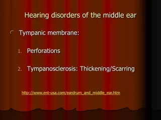
Hearing disorders of the middle ear Tympanic membrane: Perforations Tympanosclerosis: Thickening/Scarring http://www.ent-usa.com/eardrum_and_middle_ear.htm
http://www.bestforhearing.com/images/normal-ed.jpg http://www.hmc.psu.edu/ume/fcmonline/case32/images/normal-ear.jpg
TM Perforation http://www.ghorayeb.com/files/TYMPANIC_PERFORATION_LEFT_LABELED.jpg
TM Perforation http://texasearcenter.com/userfiles/Image/tmp-1.jpg
Consequences Mild hearing loss Treatment In some cases, spontaneous recovery (depends on location) Myringoplasty: Surgical reconstruction
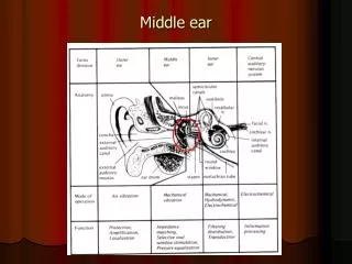
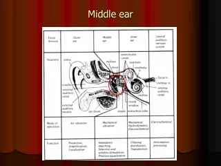
Middle ear cavity Otitis media: Very common (~70% of children in the U.S.) Infection of the mucous membrane lining the middle ear Predisposing factors: Poorly functioning Eustachian tubes Barotrauma Abnormal functioning of mucous membrane cilia Anatomical deformities Gender and demographic factors Exposure to toxic fumes
TM Retraction http://img.medscape.com/pi/emed/ckb/otolaryngology/834279-858557-860080-860208.jpg
Acute OM http://www.mdconsult.com/das/book/body/161330440-2/0/1608/f4-u1.0-B978-1-4160-2450-7..50641-1..gr3.jpg
Acute OM http://upload.wikimedia.org/wikipedia/commons/5/58/Otitis_media_entdifferenziert2.jpg
Acute OM http://knol.google.com/k/-/-/Y0fzk46N/nxV32A/AOM.jpg
Possible mechanisms Through ruptured tympanic membrane Through Eustachian tube Through blood
Characteristics of otitis media infection Two types: Acute and chronic Rapidly progressive Negative pressure in middle ear because of ET malfunction TM is retracted and appears red Pain, high temperature, pus accumulates in middle ear mucosa In severe cases, TM ruptures due to pressure. If left untreated, can progress to mastoid air cells and cause mastoiditis. http://www.ent-usa.com/eardrum_and_middle_ear.htm
Consequences Flat conductive hearing loss (degree depends on the amount of fluid) Low static compliance Type B tympanogram Absent reflexes Absent OAEs High latencies for all ABR peaks
Treatment Antibiotics Surgery Myringotomy and suction Mastoidectomy (if infection has spread to mastoid region) Tympanoplasty
PE Tubes http://www.pedisurg.com/PtEducENT/tube_in_TM.jpg
Complications of otitis media Cholesteotoma: Sac-like growth due to presence of skin in the middle ear. Dangerous, progressive, highly erosive Foul-smelling discharge (otorrhea) Treatment: Surgical removal http://www.ent-usa.com/eardrum_and_middle_ear.htm
Cholesteatoma http://chicagoear.com/med_info/images_med_info/cholesteatoma.jpg
Cholesteatoma http://www.earsurgery.org/images/Photo-31%20copy.jpg
Cholesteatoma http://my.clevelandclinic.org/PublishingImages/Head_Neck/cholesteatoma.jpg
Facial palsy • If erosion of bone occurs, facial nerve may be exposed. • Partial/Full paralysis of one side of the face. • Treatment: Surgery
Eustachian tube problems Cause: Infections, allergies, blockage due to overgrown adenoids, structural problems. Consequences: Negative middle ear pressure, retracted TM. Audiometric findings: Mild conductive hearing loss, normal static compliance, type C tympanogram.
Methods to open ET Valsalva: Close nostrils and cheek and blow out. Toynbee: Close nostrils and swallow Complications of ET malfunction: Serous effusion Mucous otitis media
Otosclerosis Causes: Hereditary in 70% of cases. Progressive in nature Higher incidence in women, adults. Clinical manifestation: Spongy bone formation over the stapes footplate. Footplate becomes fixed in the oval window.
Other clinical signs Progressive hearing loss Tinnitus Difficulty hearing while chewing Very vascular promontory, rosy glow through TM (Schwartze sign) Paracusis willisii: Speech easier to understand in the presence of background noise.
Audiometric findings Low frequency conductive hearing loss with air-bone gap. As disease progresses, hearing loss spreads to high frequencies. Bone conduction is affected, primarily at 2000 Hz (called Carhart’s notch). Type As tympanogram, absent reflexes
Treatment Earlier: Surgery to free immobilized stapes footplate. Not very successful. Fenestration (new window created in lateral semicircular canal). Effects of fenestration: ~ 25 dB hearing loss to total hearing loss, vertigo, tinnitus, poor word recognition scores, facial paralysis, repeated infections of cavity Stapes mobilization: Middle ear cavity exposed through incision in TM. Effects: Immediate improvement in hearing, however, refixation of stapes often occurred. Most successful treatment: Stapedectomy. Replaced with prosthesis Modification: Stapedotomy
Inner ear • Series of interconnecting canals or ‘labyrinths’ in the temporal bone • Two types: • Osseous • Bony • Bigger cross-sectional area • Contains fluid called perilymph • Membraneous • Soft tissue • Situated within the bony labyrinth • Contains fluid called endolymph http://research.meei.harvard.edu/Otopathology/3dmodels/download.html