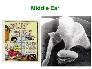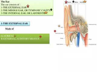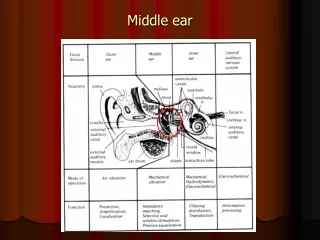
Middle Ear
Middle Ear. Differential Diagnosis of Ear Disease. External Ear. Middle Ear. Inner Ear. Cerumen impaction Auricular hematoma Perichondritis Otiis Externa Otomycosis Foreign Body External ear canal laceration -temporal bone fracture. Acute otitis media Mastoiditis

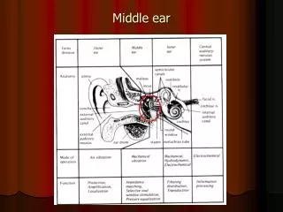
Middle Ear
E N D
Presentation Transcript
Differential Diagnosis of Ear Disease External Ear Middle Ear Inner Ear • Cerumen impaction • Auricular hematoma • Perichondritis • Otiis Externa • Otomycosis • Foreign Body • External ear canal laceration • -temporal bone fracture • Acute otitis media • Mastoiditis • Serous/chronic otitis media • Hemotympanum • Tympanic membrane perforation • Tympanic membrane retraction • Cholesteatoma • Sensorineural hearing loss • Vestibular neuritis • Meniere’s Disease • Vestibular migraine
Acute Otitis Media Peak incidence AOM is between 6 and 18 months AOM affects 40%-50% of children by age 1 By age 3 years majority (>80%) of children have had 1 episode of AOM ~ 40% of pediatric office visits in first 5 years related to otitis media ~5-10% of well visits associated with diagnosis of OME
Acute Otitis Media Diagnosis Certain diagnosis of AOM meets all 3 of the criteria: Presence of Purulent Middle Ear Effusion Rapid onset Signs and symptoms of middle-ear inflammation Otalgia No pain with pulling of ear TMJ pain Difficulty sleeping due to pain
Acute Otitis Media Diagnosis Pulling at the Ears (not reliable): Zero percent of children with ear pulling as the primary sign had an ear infection Ear pulling + fever: only 15% had ear infections Why do kids pull their ears? Itching Teething Exploration Comfort Habit Pain • Is ear pulling associated with ear infection. • Baker RB. Pediatrics. 1992 Dec;90(6):1006-7 • Diagnostic accuracy and the observation option in • acute otitis media: the Capital Region Otitis Project. • Gurnaney H, Spor D, Johnson DG, Propp R. • Int J Pediatr Otorhinolaryngol. 2004 Oct;68(10):1315-25
Acute Otitis Media Diagnosis Presence of Purulent Middle Ear Effusion Exam- Unobstructed ear canal and good light! Bulging of the tympanic membrane Limited or absent mobility of the tympanic membrane Pneumotoscopy Tympanometry Air-fluid level behind the tympanic membrane Otorrhea (purulent)
Misdiagnosis of Acute OM Over-reliance on history TM color does not predict AOME-crying makes most tympanic membranes red Failure to evaluate tympanic membrane mobility (pneumatic otoscopy) Poor light from otoscope (bulb & battery) Failure to remove cerumen Inappropriate sized speculum Lack of experience
Acute Otitis Media Treatment Why do we treat AOM? Quality of Life Suppurative Complications Once treated, when do we follow-up? If asymptomatic, follow-up is to ensure resolution of fluid This process can take up to 3 months (74%) • Intracranial Complications: • Meningitis • Extradural abscess • Subdural empyema • Lateral sinus thrombosis • Brain abscess • Otitic hydrocephalus • Extracranial Complications: • Mastoiditis • Petrositis • Facial Paralysis • Perforation of the TM • Hearing loss • CHL • SNHL • Labyrinthitis
Intracranial Complications: Meningitis Extradural abscess Subdural empyema Lateral sinus thrombosis Brain abscess Otitic hydrocephalus Extracranial Complications: Mastoiditis Petrositis Facial Paralysis Perforation of the TM Hearing loss CHL SNHL Labyrinthitis Complications of Acute OM
Acute Mastoiditis • May or may not be associated with subperiosteal abscess • Protrusion of the auricle may be secondary to osteitis of the mastoid cortex without erosion/ abscess
Acute Mastoiditis Management • IV antibiotics • Incision and drainage of subperiosteal abscess • Myringotomy and tube placement • Cortical mastoidectomy traditionally recommended
AOM vs. OME • Otitis Media with Effusion • Fluid behind TM • May result from AOM • Less sever complications • Hearing loss • Scarring/atrophy of TM • Tympanosclerosis • Do not treat with antibiotics • Ear tubes if persistent or chronic • Acute Otitis Media • Pus behind TM • Acute infection • Multiple severe complicaitons • Mastoiditis • Meningitis • Brain abscess • Facial paralysis • Treat with antibiotics • Ear tubes if recurrent
Otitis Media with Effusion Tympanic membrane characteristics Translucent or opaque Gray, white, yellow, or pink color Neutral or retracted position Reduced mobility, responds to negative pressure on pneumatic otoscopy Effusion present
Otitis Media with Effusion Treatment Intervention based on severity of hearing loss, child’s developmental status, parent preference Aggressive management of “at-risk” population Watchful waiting for at least 3 months in “non at-risk” population “Paradise Tube Article” studies only healthy, non at-risk children Nasal steroids may help Nasal decongestants/antihistamines of no proven use Antimicrobials/steroids not indicated Paradise JL., et al: Tympanostomy Tubes and Developmental Outcomes at 9 to 11 Years of Age N Engl J Med. 363 (3):248-261, 2007.
Otitis Media with Effusion Treatment Audiogram if fluid > 3 months (chronic) If normal hearing periodic re-evaluation until clear; more aggressive intervention if hearing loss, behavior problems or TM changes Surgery- Tubes with or without adenoids Tubes initially only Adenoidectomy if nasal obstruction or infection problems or if past hx of tubes Repeat surgery--adenoidectomy +/-tubes
AOM vs. OME • Otitis Media with Effusion • Fluid behind TM • May result from AOM • Less sever complications • Hearing loss • Scarring/atrophy of TM • Tympanosclerosis • Do not treat with antibiotics • Ear tubes if persistent or chronic (chronic) • Acute Otitis Media • Pus behind TM • Acute infection • Multiple severe complicaitons • Mastoiditis • Meningitis • Brain abscess • Facial paralysis • Treat with antibiotics • Ear tubes if recurrent
Types of TM Findings Serous otitis media Normal TM Mucoid Otitis Media Acute Otitis Media
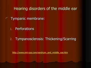
Tympanic Membrane Perforation Myringosclerosis
Tympanic Membrane Perforation • Multiple causes • Trauma (welder’s slag) • Tubes • Infection • Barotrauma • Treatment • Auidiogram • Dry ear precautions • Ciprodex for otorrhea • Tympanoplasty if no spontaneous resolution (~6 months) • 90-95% success rate • Potential complications • Otorrhea • Hearing loss • Cholesteatoma
Tympanic Membrane Retraction • Negative pressure pulls TM inward • Caused by eustachian tube dysfunction • Most likely in superior TM (“Prussack’s space”) • Loss of middle ear volume • Loss of amplification • Physical exam: • Microscope: can see retraction • Monocular otoscope: oval/angled/rotated TM • Angled/horizontal malleus • Complications: • Serous otitis media • Hearing loss • Damage to ossicles • Cholesteatoma
Cholesteatoma • Dermoid cyst in middle ear space • Recurrent otorrhea/infection in one ear • Physical exam: • Squamous debris and granulation tissue • “like a bomb went off” • May see “pearl” • Causes: • Epithelial rest (congenital) • Retraction of TM with migration of epithelium into middle ear • Perforation/trauma • Complications: • Hearing loss • Destruction of ossicles • Facial paralysis • Intracranial extension • Vertigo
Cholesteatoma • Work-up: • Audiogram • CT Temporal bones without contrast • Treatment: • Ciprodex for otorrhea • Dry ear precautions • Surgery • Mulitple surgeries required
Differential Diagnosis of Ear Disease External Ear Middle Ear Inner Ear • Cerumen impaction • Auricular hematoma • Perichondritis • Otiis Externa • Otomycosis • Foreign Body • External ear canal laceration • -temporal bone fracture • Acute otitis media • Mastoiditis • Serous/chronic otitis media • Hemotympanum • Tympanic membrane perforation • Tympanic membrane retraction • Cholesteatoma • Sensorineural hearing loss • Vestibular neuritis • Meniere’s Disease • Vestibular migraine • BPPV
Inner Ear Pathology • Key symptoms: • Sensorineural hearin gloss • Tinnitus • Vertigo • Work-up: • Audiogram • MRI of internal auditory canal with and without contrast • +/- CT temporal bones • +/-ENG • Treatment for HL • Hearing aids • Hearing aids • Hearing aids • Cochlear implant • Treatment for vertigo • Meclizine • Valium • Vestibular exercises
Summary • Ear pathology complex • Symptoms, age, type of hearing loss can help focus differential • Remember that otoscope distorts exam • Dry ear precautions always a good idea • No shame in ENT referral
Questions? Thank You!