Download
1 / 22
220 likes | 444 Vues
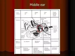
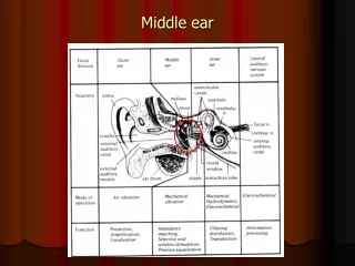
Microbiology of Middle Ear Infections. Definitions. Middle ear is the area between the tympanic membrane and the inner ear including the Eustachian tube. Otitis media ( OM ) is inflammation of the middle ear. Anatomy of the Middle Ear. OM-Classification. Acute OM Secretory ( Serous ) OM

E N D
Definitions • Middle ear is the area between the tympanic membrane and the inner ear including the Eustachian tube. • Otitis media (OM) is inflammation of the middle ear.
OM-Classification • Acute OM • Secretory ( Serous) OM • Chronic OM
OM- Epidemiology • Most common in infants 6 to 18 months of age (2/3 of cases). Improves with age, why ? • The Eustachian Tube which vents the middle ear to the nasopharynx , is horizontal in infants, difficult to drain naturally, its surface is cartilage ,and lymphatic tissue lining is an extension of adenoidal tissue from back of the nose. • Accompanied with viral URTI
OM-Pathogenesis and Risk Factors • URTI or allergic condition cause edema or inflammation of the tube. • Functions of the tube ( ventilation, protection and clearance ) disturbed. • Oxygen lost leading to negative pressure • Pathogens enter from nasopharynx into middle ear. • Colonization and infection result.
OM- Other risk factors • Anatomic abnormalities • Medical conditions such as Cleft palate ,obstruction due to adenoid or NG tube or malignancy, immune dysfunction. • Exposure to pathogens from day care. • Exposure to smoking.
OM-Microbiology-Bacterial Causes • Acute OM < 3months of age > 3 months of age • S.pneumoniae,(40%) group B Streptococcus, H.influenzae (non typable) ,Gram negative bacteria and P.aeruginosa • S.pneumoniae,H.influenzae,others eg, S.pyogenes, Moraxellacatarrhalis, S.aureus
OM-Microbiology-cont. Chronic OM Serous OM • Mixed flora in 40% of cases • P.aeruginosa, H.influenzae, S.aureus, Proteus species, K.pneumoniae, Moraxellacatarrhalis, anaerobic bacteria. • Same as chronic OM, but • Most of the effusions are sterile • Few acute inflammatory cells
OM-Viral causes • RSV -74% of viral isolates • Rhinovirus • Parainfluenza virus • Influenza virus
Clinical presentation • Acute OM Mostly Bacterial ,often a complication of viral URTI First 1-2 days: Fever (39 C), irritability, earache , muffled nose. Bulging tympanic membrane ,poor mobility and obstruction by fluid or inflammatory cells on otoscopic examination.
3-8 days: Pus and ear exudate discharge spontaneously and pain and fever begin to decrease. 2-4 weeks : Healing phase, discharge dies up and hearing becomes normal.
Serous OM • Collection of fluid within the middle ear as a result of negative pressure produced by altered eustachian tube function. • Represent a form of chronic OM or allergy-related inflammation • Tends to be chronic , with non –purulent secretions. • Cause hearing deficit.
Chronic OM • Usually result from unresolved acute infection due to in adequate treatment or host factors that perpetuate the inflammatory process. • Result in destruction of middle ear structures and significant risk of permanent hearing loss.
Diagnostic approaches of OM • Clinical examination • Tympanometry ( detect presence of fluid) • Gram stain and culture of aspirated fluid to determine the etiologic agents.
Management of OM • Acute OM requires antimicrobial therapy & careful follow up. • Antimicrobial usually empirical depending on the most likely bacterial pathogens, usually to cove S.pneumonia and H.influenzae. • Drainage of exudate may be required. • Chronic or serous OM need complex management, possibly surgical.
Complications extracranial Intracranial • Hearing loss • Tympanic membrane perforation • Mastoiditis • Cholestatoma • Labyrinthitis • others • Meningitis • Extradural abscess • Suduralempyema • Brain abscess • others