Middle ear
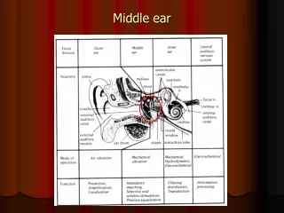
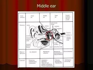
Middle ear . Middle ear structures. Middle ear cavity/tympanum Tympanic membrane Ossicles/Middle ear bones. 2-4 mm horizontal, 13 mm vertical, approximately 2 cm 3 in volume Comprised of Tympanic cavity between outer and inner ear Epitympanic recess above the TM

Middle ear
E N D
Presentation Transcript
Middle ear structures Middle ear cavity/tympanum Tympanic membrane Ossicles/Middle ear bones
2-4 mm horizontal, 13 mm vertical, approximately 2 cm3 in volume Comprised of Tympanic cavity between outer and inner ear Epitympanic recess above the TM Mastoid recess contained in the mastoid region of the temporal bone Middle ear cavity
Similar to a box with six surfaces Lateral wall or membraneous wall: Formed by TM and squamous portion of TB. Medial wall or labyrinthine wall: Promontory (outer wall of inner ear) (oval and round windows, prominence of facial nerve) Anterior wall or carotid wall (opening of ET and tendon of tensor tympani) Posterior wall or mastoid wall (tympanic aditus, fossa incudis, pyramidal prominence, facial nerve through tympanic sulcus) Inferior wall or jugular wall (tympanic plate of TB) Superior wall or tegmental wall (tegmen tympani, continuing posteriorly to tympanic atrium) Tympanic cavity
Tympanic membrane • Oval shaped lateral surface • Thick periphery called annulus with the notch of Rivinus • Located in a bony groove called tympanic sulcus • 8-9 horizontal, 9-10 mm vertical axis, 0.1 mm thick. • Held in place by fibers and cartilage • Cone shaped, translucent • 55 to 90 mm2 in area http://www.ghorayeb.com/AuricleEACAnatomy2.html
Three layers Outer: Epidermis of EAM Inner: Mucosal lining of middle ear space Middle: Collagen fibers, support for TM. Middle layer composed of two sets of fibers: • Radiates outward from center • Concentric rings of fibers • Two regions in tympanic membrane: • Superior region: Pars flaccida (defined by anterior and posterior malleolar folds and malleolar prominence) • Inferior region: Pars tensa
Tympanic membrane, cont’d. • Umbo: Region of maximum concavity • Position changes as EAM lengthens after birth • Important landmarks visible • Tympanic membrane attached to ossicles. http://hyperphysics.phy-astr.gsu.edu/hbase/sound/ear.html
Ossicles • Malleus: Hammer • Incus: Anvil • Stapes: Stirrup http://www.ghorayeb.com/StapesPics.html • Head of malleus and body of incus located in the epitympanic recess • http://oto.wustl.edu/bbears/ossicle.htm http://audilab.bmed.mcgill.ca/~daren/3Dear/mid1.html
Malleus • 9 mm long, 23-37 mg weight • Head with articular facet • Neck • Anterior and lateral processes • Handle or Manubrium
Incus • About 23-32 mg weight • Body • Short crus/process in epitympanic recess, around 5 mm • Long crus/process with lenticular process, around 7 mm (Plural of crus: Crura)
Stapes • 2-5 to 3.8 mm tall, 2.1 to 4.3 mg weight. Footplate area around 3.2 mm2 • Head, with spine for attachment of stapedial tendon • Neck • Two bony crura, anterior and posterior • Flat oval bone called footplate • Medial surface of footplate fastened to wall of oval window by annular ligament. • Unique connection allows for rocking, rather than piston-like, movement of stapes.
Ossicular connections Malleus: Manubrium of malleus connects to umbo of TM. Incus: Head of malleus connects to body of incus (incudo-mallear joint) Stapes: Inferior process of incus connects to form lenticular process, which connects to the stapes (incudo-stapedial joint) Footplate of the stapes connects to the oval window
Ossicular connections, cont’d. Suspended in the middle ear cavity by axial ligaments and muscle tendons Middle ear muscles Tensor tympani and stapedius
Tensor tympani • Enters middle ear through anterior wall of tympanum. • Connected to manubrium/neck of the malleus • Activated by the trigeminal nerve • Muscle runs parallel and superior to osseous foundation of the ET, separated by septum canalis musculotubarii) • Pulls malleus in anterior and posterior direction
Stapedius muscle • Tendon enters the middle ear space through opening in the posterior wall of the tympanum (pyramidal eminence) • Attached to the head of the stapes • Activated by the facial nerve • Pulls stapes in the posterior direction