Download
1 / 45
450 likes | 657 Vues
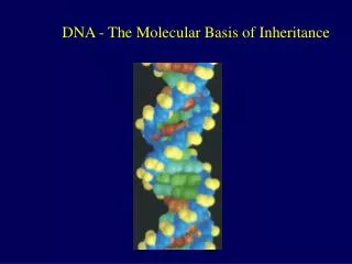
The Molecular Basis of Inheritance. Chapter 16. Life’s Operating Instructions. Hereditary information is encoded in DNA (deoxyribonucleic acid) Reproduced in all cells of the body Transmitted to offspring by chromosomes in gametes

E N D
The Molecular Basis of Inheritance Chapter 16
Life’s Operating Instructions • Hereditary information is encoded in DNA (deoxyribonucleic acid) • Reproduced in all cells of the body • Transmitted to offspring by chromosomes in gametes • DNA directs the development of biochemical, anatomical, physiological, and behavioral traits
DNA= Deoxyribonucleic acid • Nucleic Acid • Located in nucleus of cell • Genetic material inherited from parents • Genes code for specific proteins with unique code of nucleotides • Monomers= nucleotides • 3 parts to nucleotide • 5 carbon sugar (deoxyribose) • Phosphate group • Nitrogenous base
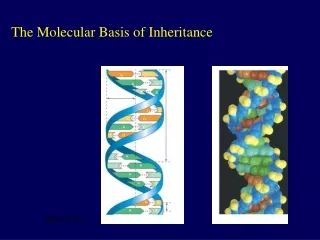
DNA • Nucleotides form polynucleotides • Double helix structure • 2 polynucleotides wrap around each other • Nitrogenous bases pair in center between 2 backbones • Strands are complementary • 4 nitrogenous bases • Adenine-Thymine • Cytosine-Guanine
DNA • Strands are anti-parallel • Two ends of strand are different from each other • One end has a phosphate attached to a 5’ carbon • Other end has a hydroxyl group attached to a 3’ carbon 5’ 3’ 5’ 3’
Chromosome Structure • Bacterial chromosome= double-stranded, circular DNA molecule associated with a small amount of protein • In bacteria, the DNA is “supercoiled” and found in a region of the cell called the nucleoid • Eukaryotic chromosomes have linear DNA molecules associated with a large amount of protein • Chromatin,a complex of DNA and protein, is found in the nucleus of eukaryotic cells
Chromosome Structure • In humans, each cell has DNA comprised of ~6 billion base pairs • Each diploid cell contains ~2 m of DNA • In total, humans contain ~100 trillion m of DNA • Enough to circle equator of Earth 2.5 million times!
Chromosome Structure • Most chromatin is loosely packed in the nucleus during interphase and condenses prior to mitosis • Loosely packed chromatin is called euchromatin • During interphase a few regions of chromatin (centromeres and telomeres) are highly condensed into heterochromatin • Dense packing of the heterochromatin makes it difficult for the cell to express genetic information coded in these regions • DNA fits into the nucleus through an elaborate, multilevel system of packing
Figure 16.22a Nucleosome(10 nm in diameter) DNA double helix(2 nm in diameter) H1 Histonetail Histones Nucleosomes, or “beads ona string” (10-nm fiber) Histones DNA, the double helix The DNA molecule binds with proteins known as histones, due to a negative charge on the strands of the DNA molecule and positive charges on histones
Figure 16.22a Nucleosome(10 nm in diameter) DNA double helix(2 nm in diameter) H1 Histonetail Histones Nucleosomes, or “beads ona string” (10-nm fiber) Histones DNA, the double helix Nucleosome is a histone complex with the DNA molecule wrapped around twice. The histone tails (amino end of protein) extend outward. The strands of DNA between the nucleosomes are called “linker DNA”.
Figure 16.22b Chromatid(700 nm) 30-nm fiber Loops Scaffold 300-nm fiber 30-nm fiber Interactions between histone tails and linker DNA result in further compaction into 30-nm fiber Replicatedchromosome(1,400 nm) Looped domains(300-nm fiber) Metaphase chromosome
Figure 16.22b Chromatid(700 nm) 30-nm fiber Loops Scaffold 300-nm fiber 30-nm fiber This fiber forms loops called looped domains attached to a protein scaffold, compacting material into 300 nm fiber Replicatedchromosome(1,400 nm) Looped domains(300-nm fiber) Metaphase chromosome
Figure 16.22b Chromatid(700 nm) 30-nm fiber Loops Scaffold 300-nm fiber 30-nm fiber The looped domains condense further into the chromosomes visible during the stages of mitosis Replicatedchromosome(1,400 nm) Looped domains(300-nm fiber) Metaphase chromosome
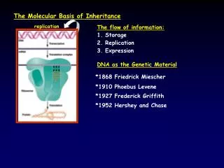
Genetic Material • Early in the 20th century, the identification of the molecules of inheritance loomed as a major challenge to biologists • T. H. Morgan’s group showed genes are located on chromosomes • 2 components of chromosomes—DNA and protein—became candidates for the genetic material
Genetic Material • Frederick Griffith (1928) • Experiments with two strains of a bacteria causing pneumonia • one pathogenic and one harmless • When he mixed heat-killed remains of the pathogenic strain with living cells of the harmless strain, some living cells became pathogenic • Transformation= a change in genotype and phenotype due to assimilation of foreign DNA
Genetic Material • Studies in 1944 by Oswald Avery, Maclyn McCarty, and Colin MacLeod provided experimental evidence that only DNA worked in transforming harmless bacteria into pathogenic bacteria • In 1950, Erwin Chargaff reported that DNA composition varies from one species to the next • Made DNA a more credible candidate for the genetic material
Genetic Material • At this time, it was known that DNA is a polymer of nucleotides, each consisting of a nitrogenous base, a sugar, and a phosphate group • Twofindings became known as Chargaff’s rules • The base composition of DNA varies between species • In any species the number of A and T bases are equal and the number of G and C bases are equal
Genetic Material • More evidence for DNA as the genetic material came from studies of viruses that infect bacteria • Such viruses, called bacteriophages (or phages), are widely used in molecular genetics research • In 1952, Alfred Hershey and Martha Chase designed an experiment using a phage known as T2 and E. coli cells
Figure 16.4-3 EXPERIMENT Emptyproteinshell Radioactiveprotein Radioactivity(phage protein)in liquid Phage Bacterial cell Batch 1:Radioactivesulfur(35S) DNA PhageDNA Centrifuge Pellet (bacterialcells and contents) RadioactiveDNA Batch 2:Radioactivephosphorus(32P) Centrifuge Radioactivity(phage DNA)in pellet Pellet
Genetic Material • Experiment with T2 and E. coli cells • Results showed only one of the two components of T2 (DNA or protein) enters an E. coli cell during infection • Concluded that the injected DNA of the phage provides the genetic information
DNA • After DNA was accepted as the genetic material, the challenge was to determine how its structure accounts for its role in heredity • Maurice Wilkins and Rosalind Franklin were using a technique called X-ray crystallography to study molecular structure • Franklin produced a picture of the DNA molecule using this technique
DNA • Franklin’s picture was used by James Watson and Francis Crick to model the structure of DNA • DNA was helical • Width of the helix and spacing of nitrogenous bases • The pattern in the photo suggested that the DNA molecule was made up of two strands, forming a double helix
(a) Space-fillingmodel Key features ofDNA structure (c) Nitrogenous bases Figure 16.7 5 end G C Hydrogen bond C G 3 end C G C A T G 3.4 nm A T C G C G C G A T 1 nm G C T A C G C G T A G C 3 end A T A T 0.34 nm 5 end A T (b) Partial chemical structure Sugar-phosphate backbone Strands are anti-parallel
Base Pairing Purine purine: too wide Pyrimidine pyrimidine: too narrow Purine pyrimidine: widthconsistent with X-ray data • Watson and Crick reasoned that the pairing was more specific • Adenine (A) paired only with thymine (T) and guanine (G) paired only with cytosine (C) • The Watson-Crick model explains Chargaff’s rules: in any organism the amount of A = T, and the amount of G = C
Sugar Sugar Adenine (A) Thymine (T) Sugar Sugar Guanine (G) Cytosine (C)
Form=Function • Watson and Crick noted that the specific base pairing suggested a possible copying mechanism for genetic material • Since the two strands of DNA are complementary, each strand acts as a template for building a new strand in replication • In DNA replication, the parent molecule unwinds, and two new daughter strands are built based on base-pairing rules
“Daughter” DNA molecules,each consisting of oneparental strand and onenew strand Separation ofstrands (b) (c) • Watson and Crick’s semiconservative model of replication • After replication, each daughter molecule will have one old strand (from the parent molecule) and one newly made strand A A A T T T T A G C C G G G C C A T A A A T T T T A T A A T T A C C G G G C C G (a) Parent molecule • Competing models were the conservative model (the two parent strands rejoin) and the dispersive model (each strand is a mix of old and new) • Rejected by later experiments by Matthew Meselson and Franklin Stahl
DNA Replication • Replication begins at origins of replication, where the two DNA strands are separated, opening up a replication “bubble” • Bacterial DNA has one origin of replication for its circular DNA • A eukaryotic chromosome may have hundreds or even thousands of origins of replication, increasing speed of replication • Replication proceeds in both directions from each origin, until the entire molecule is copied • At the end of each replication bubble is a replication fork, a Y-shaped region where new DNA strands are elongating
DNA Replication: Duplicating the code At the replication forks, both strands replicated at same time in the 5’ to 3’ direction
DNA Replication: Duplicating the code Helicase- unwinds double helix
DNA Replication: Duplicating the code Topoisomerase- prevents overwinding at replication fork by breaking, swiveling, and rejoining DNA strands
DNA Replication: Duplicating the code Single-strand binding proteins bind to and stabilize single-stranded DNA
DNA Replication: Duplicating the code DNA primase- start an RNA chain from scratch and adds RNA nucleotides one at a time using the parental DNA as a template
DNA Replication: Duplicating the code RNA primer- short (5–10 nucleotides long) RNA molecule that serves as the starting point for the new DNA strand
DNA Replication: Duplicating the code DNA polymerases- catalyze the elongation of new DNA at a replication fork by adding nucleotides only to the free 3’ end of a growing strand
Figure 16.14 Template strand New strand 5 5 3 3 Sugar A T A T Base Phosphate C G C G C G G C DNApolymerase OH 3 A T A T OH P P P i P P 3 C C Pyrophosphate OH 2P i Nucleosidetriphosphate 5 5
DNA Replication: Duplicating the code Leading strand: Template strand is 3’ to 5’ Lagging strand: Template strand is 5’ to 3’
DNA Replication: Duplicating the code DNA polymerase synthesizes the leading strand continuously, moving toward the replication fork in 5’ to 3’ direction
DNA Replication: Duplicating the code To elongate the lagging strand, DNA polymerase must work in the direction away from the replication fork
DNA Replication: Duplicating the code Lagging Strand made by Okazaki fragments -small sections of DNA made in 5’ to 3’ direction
DNA Replication: Duplicating the code DNA ligase- joins the Okazaki fragments
Preventing Mistakes • DNA polymerases proofread newly made DNA, replacing any incorrect nucleotides • Mismatch repair= repair enzymes correct errors in base pairing • Nucleotide excision repair= a nuclease cuts out and replaces damaged stretches of DNA • Error rate after proofreading repair is low but not zero • Sequence changes may become permanent and can be passed on to the next generation • These mutations are the source of the genetic variation upon which natural selection operates
Replicating the Ends of DNA Molecules • Replication mechanism in eukaryotic cells provides no way to complete the 5 ends • Repeated rounds of replication produce shorter DNA molecules with uneven ends • Not a problem for prokaryotes with circular chromosomes • Eukaryotic chromosomal DNA molecules have special nucleotide sequences at their ends called telomeres • Repetitive DNA sequences • Telomeres do not prevent the shortening of DNA molecules, but they do postpone the erosion of genes near the ends of DNA molecules • It has been proposed that the shortening of telomeres is connected to aging
Replicating the Ends of DNA Molecules • If chromosomes of germ cells became shorter in every cell cycle, essential genes would eventually be missing from the gametes they produce • An enzyme called telomerase catalyzes the lengthening of telomeres in germ cells, preventing this problem • The shortening of telomeres might protect cells from cancerous growth by limiting the number of cell divisions • There is evidence of telomerase activity in cancer cells, which may allow cancer cells to persist