Optimizing Oxygenation and Perfusion: Essential Concepts for Respiratory Health
310 likes | 602 Vues
This educational guide discusses the crucial factors affecting oxygenation and perfusion in the human body, focusing on the respiratory and cardiovascular systems. It covers the anatomy of lungs, gas exchange processes, respiratory gas transport, cardiovascular function, and factors impacting cardiopulmonary functioning. The guide also explores developmental considerations, alterations in health, medications, and lifestyle factors that can influence respiratory health. Nursing interventions for promoting optimal respiratory functioning and improving breathing techniques are outlined to enhance patient care and well-being.

Optimizing Oxygenation and Perfusion: Essential Concepts for Respiratory Health
E N D
Presentation Transcript
Oxygenation and Perfusion depend on….. • The condition of the airway system from nose to alveoli • Properly functioning alveolar system in lungs to facilitate gas exchange • Properly functioning cardiovascular and hematologic systems that carry nutrients and wastes to and from body cells
Function of the Airway • Warm, filter, humidify inspired air • Movement of air • Clearance of mucus by cilia • Production of pulmonary surfactant
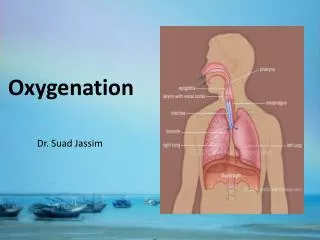
Anatomy of the Lungs • Main organs of respiration • Extend from the base of the diaphragm to the apex above the first rib • The right lung has three lobes; the left lung has two. • The lungs are composed of elastic tissue (alveoli, surfactant, pleura).
Pulmonary Ventilation • Inspiration: the active phase of ventilation • Involves movement of muscles and the thorax to bring air into the lungs • Expiration: the passive phase of ventilation • Movement of air out of the lungs
Process of Ventilation • Inspiration begins when the diaphragm contracts and descends, lengthening the thoracic cavity. • The external intercostal muscles contract, lifting the ribs upward and outward. • The sternum is pushed forward, enlarging the chest from front to back • The relaxation of these structures results in expiration.
Gas Exchange (Respiration) • Refers to the intake of oxygen and release of carbon dioxide • Made possible by respiration and perfusion • Gas exchange occurs via diffusion at the alveolar level • Alveoli have a single layer membrane that allows the exchange of oxygen and carbon dioxide between the alveolar sacs and the capillaries
Transport of Respiratory Gases • Oxygen is carried in the body via plasma and red blood cells. • Oxygen and carbon dioxide are carried by red blood cells in the form of oxyhemoglobin and carboxyhemoglobin. • Internal respiration occurs between the circulating blood and tissue cells in the body. This is called perfusion.
Cardiovascular System • Vital for exchange of gases • Composed of the heart and the blood vessels • The heart is a cone shaped, muscular pump, divided into four hollow chambers: • Atria receive blood from the veins: superior and inferior vena cava (from body) and the left and right pulmonary veins (from lungs). • Ventricles force blood out of the heart through the arteries: aorta (to body) and the left and right pulmonary arteries (to lungs).
Factors Affecting Cardiopulmonary Functioning and Oxygenation • Developmental considerations • Alterations in health • Medications • Lifestyle
Developmental Considerations • Infants are nose-breathers • Children have shorter airways • Older adults have decreased elasticity in lungs and CV system and skeletal changes that can affect lung performance
Alterations in Health Factors • Cardiac disease • Vessel disease • Blood clots • Pulmonary disease • Skeletal abnormalities • Diabetes • Deconditioning and bedridden • Swallowing difficulties • Depression • Trauma • Infections
Alterations in Respiratory Function • Hypoxia: inadequate amount of oxygen available to the cells • Dyspnea: difficulty breathing • Hypoventilation: decreased rate or depth of air movement into the lungs
Alterations in the Cardiovascular System • Dysrhythmia (irregularity) • Myocardial ischemia (meaning low oxygen and perfusion to the heart muscle) • Angina (pain from severe ischemia) • Myocardial infarction (death of tissue from prolonged ischemia) • Heart failure (damaged pump)
Medication factors • Cardiac medications—can control irregularities, improve pump function, control pressures, treat or prevent pain • Anticoagulants and aspirin • Pulmonary medications—can open airways • Antibiotics—treat infection in the respiratory tract • Narcotics—can slow respiratory rate and decrease air exchange; however morphine is a vasodilator • Herbals
Lifestyle Factors • Smoking • Sedentary • Illegal drugs • Dietary choices • Occupational exposure • Geographic location
Nursing Interventions Promoting Adequate Respiratory Functioning • Effective breathing techniques • Effective coughing techniques • Suctioning the airway • Meeting oxygenation needs with medications • Smoking cessation • Avoiding environmental triggers
Promoting Effective Breathing • Deep breathing • Using incentive spirometry (IS) • Pursed-lip breathing • Diaphragmatic breathing • Positioning
Promoting Comfort • Positioning • Maintaining adequate fluid intake • Providing humidified air • Performing chest physiotherapy • Maintaining good nutrition • Pacing physical activities
Medications • Cough suppressants • Expectorants • Lozenges • Bronchodilators • Antiinflammatories • Antibiotics • Oxygen
Administering Inhaled Medications • Bronchodilators: open narrowed airways. Can be given po, IV, or by inhalation. • Nebulizers: disperse fine particles of liquid medication into the deeper passages of the respiratory tract (breathing treatment to dilate bronchioles) • Meter-dose inhalers: deliver a controlled dose of medication with each compression of the canister. Can be rescue or maintenance. • Dry powder inhalers: breath-activated delivery of medications
Common Oxygen Delivery Systems • Nasal cannula—1-6 L/min. Can be humidified. • Venturi mask—4-10 L/min. O2 mixes with room air, CO2 is expelled. Has large tube attached. Allows for the most accurate delivery of whatever percentage the HCP wants. • Simple mask—6-8 L/min. O2 mixes with room air, CO2 is expelled • Partial rebreather mask—8-11 L/min. Has reservoir bag that traps exhaled CO2. O2 mixes with small amount of trapped expelled CO2 on inspiration. • Nonrebreather mask—12 L/min. Has reservoir bag that holds oxygen. Highest delivery of O2. No mixing occurs because CO2 is exhaled through vents. • Tent—used for peds. Plastic cover over bed. Motor keeps air cool and humidified. Side vents allow nsg care. Can cause damp linens and clothing.
Precautions for Oxygen Administration • Avoid open flames in the patient’s room. • Place “no smoking” signs in conspicuous places. • Check to see that electrical equipment in the room is in good working order. • Avoid wearing and using synthetic fabrics (builds up static electricity). • Avoid using oils in the area (oils ignite spontaneously in oxygen).
Type of Artificial Airways • Oropharyngeal and nasopharyngeal airway-to maintain airway patency and facilitate suctioning • Endotracheal tube-to deliver oxygen and medications when patient is unconscious • Tracheostomy tube-inserted directly into the trachea from the outside to maintain airway patency when upper airway is obstructed
Two Types of Tracheostomy Sets: Cuffless and Cuffed (Cuffed allows ventilator)