Optic chasm
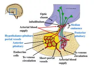
Optic chasm. infudibulum. Median eminence. Arterial blood supply. Posterior pituitary. Hypothalamo-pituitary portal vessels. Anterior pituitary. Endocrine cells. To venous circulation. To venous circulation. Short portal vessels. Arterial blood supply. Axon to primary

Optic chasm
E N D
Presentation Transcript
Optic chasm infudibulum Median eminence Arterial blood supply Posterior pituitary Hypothalamo-pituitary portal vessels Anterior pituitary Endocrine cells To venous circulation To venous circulation Short portal vessels Arterial blood supply
Axon to primary capillaries Primary capillaries Median eminence Superior hypophyseal artery Pituitary stalk Portal venules Posterior pituitary Anterior pituitary Secondary capillaries
Transduction Pathways of Releasing Hormones Pathway Hormone Location of action PKAa PIb Corticotrope of anterior pituitary (ACTH) + CRH Thyrotrope of anterior pituitary (TSH) +c TRH Gonadotrope of anterior pituitary (LH & FSH) GnRH AVP Corticotrope of anterior pituitary; assists CRH in releasing ACTH +
Transduction Pathways of Releasing Hormones (cont.) Pathway Hormone Location of action PKAa PIb GHRH Somatomammotrope of anterior pituitary (GH) (?) + Somatomammotrope of anterior pituitary (inhibits GH release) Somatostatin +d PRL Release inhibitor aPKA, protein kinase A bPI, phosphatidylinositol pathway cThe increase of cytoplasmic calcium concentration may be important in the actions of PI stimulation dInhibitory pathway of PKA
Effects of hypophysectomy • 1. Cessation of growth & the retention of juvenile features. • 2. Atrophy of adrenal cortes (zona fasciculata). • 3. Atrophy of thyroid. • 4. Decreased gonadal function in the adult. • 5. Alterations in the metabolism of lipids, proteins, and carbohydrates. • 6. Blanching of pigment cells in the skin of the lower vertebrates (fishes, amphibians, & reptiles
I. Chemistry Straight chain polypeptide of 191 AA (Two S-S bonds) Mole Weight: 22,000 deltons Produced by somatotroph (acidophil) cells Plasma Concentration 3-10 ng/ml Circulating half-life: 20-30 minutes Broken down by the liver Growth Hormone (GH)Somatotropin
II. Biologic Actions A. Supports Osteogenesis (Epiphyseal-diaphyseal plate) Stimulates release of peptide somatomedin from the liver 1. Oversecretion (acidophilic adenoma) (a) Giantism (b) Acromegaly 2. Undersecretion (a) Pituitary dwarf (b) Simmond’s disease (hypophyseal cachexia)
Facial changes in a patient with acromegaly. Panel at left shows appearance of normal young woman. Middle & right show effects of acromegaly with coarsening of facial features.
Hand of someone with acromegaly (left) placed next to normal hand (right).
Somatotropin (cont.) • B. Promotes protein synthesis (anabolic effect) • Retards AA catabolism: transport into cell. • AA retention causes positive nitrogen - phosphate balance. Na+ + K+ excretion • C. Diabetogenic effect (Houssay animal) • Blocks hexokinase
Somatotropin (cont.) • Affects carbohydrate metabolism by: • 1. hyperglycemia • 2. Inhibiting insulin action • 3. muscle glycogen • 4. Production of permanent diabetes mellitus (destroys B cells)
Somatotropin (cont.) • D. Peripheral mobilization of fats • (Pharmacologic doses) • [serum fatty acids] • E. Increase intestinal absorption of calcium • F. Increase renal tubular reabsorption of phosphorus
Somatotropin (cont.) • III. Regulation of release • A. Stimulation (via Somatotropin Releasing Hormone) SRH • Deficiency of energy substrate: • 1. Hypoglycemia • 2. Exercise • 3. Fasting • 4. Plasma protein • 5. Sleep
Somatotropin (cont.) • B. Inhibition (via somatostatin) IH • 1. Serum glucose • 2. Cortisol • 3. REM sleep
Somatotropin (cont.) • IV. Potential Clinical Use for Hypothalmic Hormones • A. Somatotropin Releasing Hormone • Treats GH deficiency effects-short stature • B. Somatostatin (14 AA peptide) • Treatment of acromegaly
Growth Curve 100 80 % of total growth 60 40 20 0 4 8 12 16 20 Age (years)
1.2 1.1 • • • 1.0 • • 0.9 • 0.8 Somatomedin Level (U/mL) 0.7 • 0.6 0.5 • 0.4 (8) (6) (6) (4) (5) (5) (2) 2 4 6 8 10 12 14 0 Adult Age (years)
P P P Growth H. + thyroxine Growth H. Wt. throxine control Age
Prolactin Chemistry Unbranched polypeptide of 198 AA (human). It has 3 disulfide bridges and a mole wt. of ~ 25,000. Secreted by acidphil cells (lactotrophs) of the adenohypophysis. The “prolactins” have a myriad of effects among vertebrates and hence a myriad of different names (lactogenic hormone; mammotrophin; galactin; luteotrophin; etc.) The “N&C” terminals are similar to those in growth hormone. Half-life of 15-25 minutes Serum level 8 ng/mL female : 5 ng/mL male
Prolactin (cont.) • Biologic Actions • A. Non-Humans • 1. Luteotropic effect (rats)- maintenance of the functional activity of the corpus luteum & release of progesterone (controlled by LH in humans.) • 2.Maternal behavior (rabbits)- injections of prolactin into non-pregnant rabbits results in nest building.
Ovine (sheep) prolactin. The black bars indicate disulfide bridges.
Prolactin (cont.) • B. Human Functions • 1. Mammotrophic effect- stimulates the mammary epithelium to secrete milk (i.e. produce milk.) • 2. Affects function of adrenals, gonads, steroid synthesis, and lipolysis.
Prolactin (cont.) • Regulation of Release • A. Secretion (PRH- Prolactin Releasing Hormone) • 1. Pregnancy- reaches peak during parturition • 2. Stimulation of nipples (nursing baby) • 3. Surgical or psychologic stress • 4. Coitus
Prolactin (cont.) • B. Inhibition (PIH- Dopamine) • 1. Dopmine is a product of L-Dopa (used to treat Parkinson’s disease- neurological disorder- tremors) • 2. Estrogen & progesterone- high levels during pregnancy inhibit action of prolactin. After birth, milk production increases.
Causes of Increased Concentration of Production in Serum Pregnancy Pituitary tumors Various drugs (e.g., chlorpromazine) Physiological Pathological Pharmacological Stress (e.g., surgery) Pituitary stalk sections Sleep Oral contraceptives Hypothalmic disorders, Sucking e.g., sarcoid infiltration Estrogens Exercise Chiari-Frommel syndrome Thyrotropin-releasing hormone Sexual intercourse Renal failure Insulin-induced hypoglycemia Hypothyroidism Arginine Nelson’s syndrome Adrenal insufficiency Ectopic production by tumors Parathormone
CorticotropinAdrenocorticotrophic Hormone (ACTH) Chemistry Straight chain polypeptide of 39 AA & a mole weight of 4,500. It is secreted by corticotroph cells (basophil). ACTH has a 13 AA sequence from its N-terminal which is identical with MSH. It therefore has a natural tendency to melanosize cells like B-lipotrophin (AA 41-58) Biologic Actions 1. ACTH controls the release of glucocorticoids (cortisol and corticosterone) from the zona fasciculata of the adrenal cortex. 2. Release of adrenal androgens
Zona Glomerulosa Zona Fasciculata Medulla Zona Reticularis
Regulation of Release • Almost any type of physical or mental stress • causes the release of CRH from the hypoth- • alamus. This causes the release of ACTH • which glucocorticods.
Regulation of Release (cont.) • BIOASSAYS • 1. Depletion of ascorbic acid (vitamin C). • a) formation of collagen • b) [iron] in body fluids • c) Scurvy (20-30 week deficiency) • Failure of wounds to heal ( collagen) • Cessation of bone growth • Fragile walls of blood vessels
(cont.) • 2. Depletion of cholesterol. • 3. Incubation methods: • Incubate adrenal slices with ACTH. • Measure cortisones produced. • 4. Maintenance of adrenal weight in • animals.
Thyrotropin • I. CHEMISTRY: • TSH, FSH, & LH are glycoproteins produced by basophil cells and having • chemically dissimilar subunits non-covalently linked together.
Thyrotropin (cont.) • Subunits: • a) Alpha - this subunit is identical in the • identical in the three hormones. • b) Beta - provides hormonal specificity. • Produced by thyrotroph (basophil) cells • Mole wt. 26,000 • Circulating half-life is 60 minutes • Broken down by kidney
Thyrotropin (cont.) • II. BIOLOGIC ACTIONS • 1. Maintenance of the structural and • functional integreties of the thyroid • gland. • 2. Controls iodine uptake (iodide pump) • 3. Maintains normal secretory epithelium- • low columnar.
Thyrotropin (cont.) • 4. Causes production and release of • thyroxine.
Thyroid Follicles Hypoactive Hyperactive (hyperthyroidism) Normal depleted colloid colloid secretory epithelium
Thyrotropin (cont.) • III. Regulation of release • A. Stimulation (via thyrotropin releasing hormone) • Negative feedback • 1) thyroxine • 2) body temperature • B. Inhibition • 1) serum thyroxine • 2) body temperature
Thyrotropin (cont.) • IV. Assay Methods • A. Bioassay • 1) height of secretory epithelium • 2) Number of colloid droplets in cells • 3) Iodine depletion in 1-day old chicks • 4) Uptake of radioactive iodine • B. Radioimmunoassay
FolliculotropinFollicle Stimulating Hormone (FSH) • Chemistry • Same as Thyrotropin (produced by gonadotroph cells) • Alpha= 92 AA • Beta= 118 AA • Biologic Action • A. Female • 1. Stimulates young ovarian follicles to develop multiple layers of granulosa cells & form antra.
Folliculotropin (cont.) • 2. Stimulate production of estrogen by developing follicle • B. Male • Stimulates the seminiferous tubules- spermatogenesis • Regulation of release • Concentration of circulating estrogens- negative feedback. • Note: Both estrogen production & completed spermatogenesis require LH.
OOGONIA NUMBERS(Text P. 630) • 5 months of gestation = 7 million • Birth = 2 million • Puberty = 300,000 to 400,000 • Released during sexual maturity (40 years) = 480
Germinal epithelium Corpus albicans Corpus luteum Time Ovulation Primary follicle Growing primary follicle Primary oocyte Corona radiata Secondary oocyte Zona pellucida Cumulus oophorous Secondary follicle Time Antrum Graafian follicle
ESTROGENS (17 BETA ESTRADIOL) • Biologic Actions • 1. Act as “growth hormones” stimulating mitosis in the mammary glands and the female reproductive system (uterus and vagina).
ESTROGENS (cont.) • 2. Promote the deposition of fat in the breast, thighs and buttocks, thereby decreasing the specific gravity in females. • 3. Promote the early closing of the growth • plates.
Spermatic cord Efferent ductules epididymis Rete testis within mediastinum testis testis Seminiferous tubules
Lumen of seminiferous tubule Basement membrane Interstitial cells Germinal epithelial cells Spermatazoa
LutotropinInterstitial Cell StimulatingHormone (ICSH) • Chemistry • Same as Thyrotropin • Half-life of one hour • Alpha=96 AA • Beta=120 AA
Lutotropin (cont.) • Biologic Action • A. Female • 1. Release of estrogen from developing follicle. • 2. Promotes ovulation. Ovulatory surge in concentration just before ovulation. • 3. Affects luteinization of ruptured follicle. • 4. Causes release of progesterone from corpus luteum
Lutotropin (cont.) • Biologic Action • B. Male • 1. Affects spermatogenesis- completes process. • 2. Stimulates Leydig cells to produce testosterone. • Regulation of Release • Negative feedback with progesterone • Note: This is why HCG is needed during pregnancy