Brain Research Methods.
Brain Research Methods. By Rachel Tierney. Direct Brain Stimulation Techniques. Direct brain stimulation consists of a device that delivers a weak electrical impulse to disrupt neurons in specific areas of the brain whilst the patient is fully aware and conscious.

Brain Research Methods.
E N D
Presentation Transcript
Brain Research Methods. By Rachel Tierney.
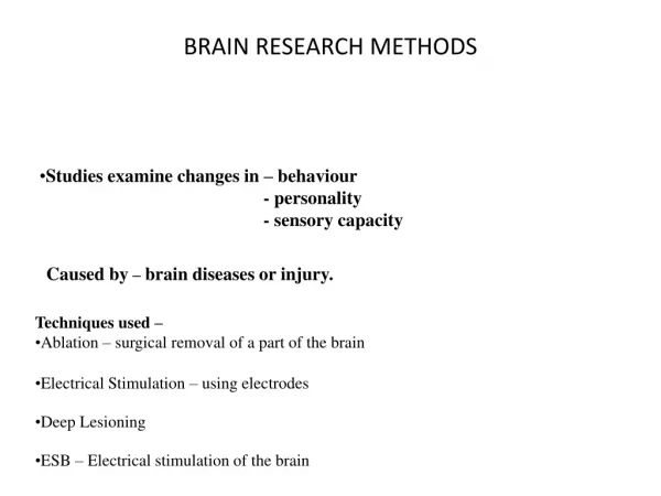
Direct Brain Stimulation Techniques. • Direct brain stimulation consists of a device that delivers a weak electrical impulse to disrupt neurons in specific areas of the brain whilst the patient is fully aware and conscious. • Two types – Electrode stimulation and Transcranial Magnetic Stimulation (TMS). • Direct brain stimulation techniques only identify the function of the brain.
Electrode Stimulation. • Wilder Penfield’s research in the 1940’s to 1960’s established that when certain areas of the brain were stimulated by electric currents a part of the patient’s body would react. He then used this information to help treat epileptic patients. • http://www.youtube.com/watch?v=kNdM9JhTPJw An electrode is a small fine wire that can be placed onto or inserted into specific areas of the brain.
Electrode Stimulation Advantages. Limitations. Extremely invasive. Considered unacceptable with today’s ethical standards. Difficulty in generalising results as most studies were done on epileptic patients with not normally functioning brains. • Helped create a ‘map’ of the brain. • Provided a way for researchers to identify the locations and functions of brain areas, including hemispheric specialisation. • Generally very consistent results
Transcranial Magnetic Stimulation (TMS) • TMS is a direct brain stimulation technique that involves directing a magnetic field pulse through the skull and then disrupts neuron activity in specific areas of the cerebral cortex. • Single pulse TMS & Repetitive TMS (rTMS) • rTMS induces consecutive impulses that cause neurons to lose their ability to fire.
Transcranial Magnetic Stimulation (TMS) Advantages Limitations Can’t be used on patients with any metal or implanted medical devices in their body. rTMS may induce scalp pain or mild headaches. Very rare cases of inducing seizures. Only penetrates the part of the brain directly below the skull. • Non–invasive. • Completely safe and harmless. • Allows researchers to conduct research ethically on human participants. • Provides information about function of brain.
Electroencephalograph (EEG) • Electrodes are placed on the scalp(cleaned) in numerous positions to identify the neuron activity in different areas of the cerebral cortex. • Detects, amplifies and records electrical brain activity.
Electroencephalograph (EEG) Advantages Limitations Only provides general information. Difficult to identify precise areas of brain activity. The skull weakens the electrical activity. • Non-invasive and harmless. • Extremely cost effective compared to other methods. • Provides information about the function of the brain even whilst the participant is doing long tasks.
Structural Neuroimaging Techniques. • Structural neuroimaging techniques can capture detailed images of the brain structure and anatomy. • Two types – CT(computerised tomography) & basic MRI(magnetic resonance imaging)
Computerised Tomography (CT) • A.K.A – Computerised axial tomography (CAT). • This technique uses computer technology to take snapshots of the brain which is separated into ‘slices’. • An injection of iodine called contrast is injected to highlight the blood vessels in the brain. • The patient must lie very still as their head is inserted into the scanner which rotates to take pictures from different angles. http://www.youtube.com/watch?v=M-4o0DxBgZk – Apply this to the brain instead of the body!
Computerised Tomography (CT) Advantages Limitations Very expensive procedure Does not show functionality of the brain on the structure. • Rarely has side effects and is non-invasive. • Shows brain structure. • Useful to identify location and size of tumours.
Magnetic Resonance Imaging (MRI) • Is a structural neuroimaging technique similar to the CT that uses magnetic fields and radio waves to vibrate atoms in neurons to project an image of the brain. • Colour is used on the image to identify highly active neurons which resemble high brain activity and vice versa.
Magnetic Resonance Imaging (MRI) Advantages Limitations Only shows structure. People with metal implants in their body cannot use it. • Clearer, more sensitive and more detailed than a CT. • Non-invasive and harmless. • Almost photographic quality images. • Does not use radioactive substances or X-rays.
Functional Neuroimaging Techniques. • These are techniques that can identify not only the brain structure like the CT and MRI can but also they have the ability to show the functions of the brain ‘at work’. • Three types – PET(Positron Emission Tomography), SPECT(single photon emission computed tomography) and fMRI(functional magnetic resonance imaging).
Positron Emission Tomography (PET) • PET uses radioactive tracer which is either injected or consumed to track blood flow through measuring how much glucose is being used by neurons in a particular part of the brain. • A computer-generated image shows the brain ‘at work’.
Positron Emission Tomography (PET) Advantages Limitations PET scans are less detailed than MRI scans. Radioactivity decays rapidly. Doesn’t always pick up rapid changes in brain activity due to 40 second intervals. • Can clearly show brain tumours, PET is more sensitive than CT and MRI. • Enables researchers to establish the role of the brain in behaviour and mental processes. • Shows function and structure. http://www.youtube.com/watch?v=5KXIDUo18aA
Single Photon Emission Computed Tomography (SPECT) • SPECT is very similar to PET but it uses a longer lasting radioactive tracer. • A scanner and computer construct either two or three dimensional images of the brain activity.
Single Photon Emission Computed Tomography (SPECT) Advantages Limitations Images are not as good as PET and have low resolution. • Longer lasting radioactive tracer can detect brain activity on longer tasks. • Shows both function and structure. • Non-invasive. • Significantly less expensive than PET.
Functional Magnetic Resonance Imaging (fMRI) • fMRI machines use oxygen levels in the blood of the brain to determine the activity of certain areas when the brain is ‘at work’. • fMRI can take rapid pictures of the brain and therefore capture more brain activity and changes.
Functional Magnetic Resonance Imaging (fMRI) Advantages Limitations Levels of activity may not be related to the tasks that they are asked to complete. Costly. • Does not require radioactive substances. • Colour coding and detailed images make it relatively easy to read.