Download
1 / 28
280 likes | 366 Vues
Explore a case of pediatric stroke following minor head trauma in a 9-year-old boy presenting with ataxia and speech difficulties. Learn about differential diagnosis, work-up, and therapeutic approaches.

E N D
Edward P. Sloan, MD, MPHAssociate ProfessorDepartment of Emergency MedicineUniversity of Illinois College of MedicineChicago, IL
Attending PhysicianEmergency MedicineUniversity of Illinois HospitalOur Lady of the Resurrection HospitalChicago, IL
Global Objectives • Improve care of the neuro patient • Minimize morbidity and mortality • Expedite disposition • Optimize resource utilization • Enhance our job satisfaction • Maximize Rx options, success
Sessions Objectives • Review a case of apparent stroke • Understand how stroke can complicate minor brain injury in children • Examine how practice in the ED can optimize outcome in these pediatric patients
Case Presentation… A 9 year old young man was brought to the ED because he was “walking like he was drunk”, with speech difficulties and illegible hand writing. He had had these symptoms for over two days, and his pediatrician had obtained a plain brain CT, which was negative. He had no headache nor any visual changes.
Case Presentation… The child had fallen while riding his bike 12 days earlier, landing on his face, without LOC, as witnessed by his siblings. His mother saw him 30 minutes later, and noted him to be “ a little dazed”, but otherwise OK. He had amnesia to the event, and complained of a diffuse headache for three days following the accident. His history was otherwise not contiributory.
Case Presentation… On physical exam, he had some resolving abrasions and ecchymosis of the right malar eminence. Although he comprehended speech adequately, his own speech was sparse. He had diffuse weakness (4/5) of all extremities, and slightly hyperreflexia on his R side, including a positive Babinski’s reflex on the R. He was noted to have cerebellar ataxia as noted on finger-to-nose and heel-to-shin movements of the R extremities. He had a moderately ataxic gait in the ED, especially with heel-to-toe walking.
Clinical Questions What is your Differential Dx? What work-up would you do? What therapies would you provide?
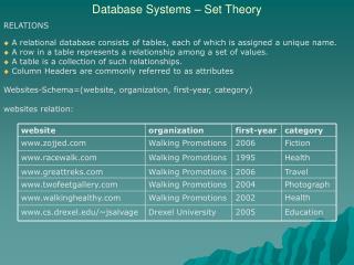
Pediatric Stroke Hemorrhagic strokes > ischemic 1.9 per 100,000 per year (vs. 1.2) Ischemic strokes due to arterial vascular or venous occlusion
Pediatric Ischemic Stroke Thromboembolism Vascular arterial occlusion Sinovenous thrombosis Occlusion of venous sinuses or cerebral veins
Head Injury and Peds Stroke Many case series and reports In series, 20-60% due to injury Often minor head trauma Often > 24 hours before sx onset
Clinical Presentation Hemiparesis, speech abn, ataxia Transient blindness No large soft tissue injury Often no skull fracture evident
ED Diagnosis in Peds Stroke Non-infused CT best first test Detects hemorrhage Will miss ischemic stroke signs DWI MRI to detect subtle changes MR angiography for vascular abn
In-Hospital Evaluation Angiography or MRA LP Serum testing Echocardiogram Coagulation testing
Non-Trauma Etiologies CNS vascular abnormalities Vasculitides Infectious causes Cardiac causes Hyper-coagulable states Demyelinating diseases Idiopathic ischemic infarct??
Ongoing Stroke Therapy German study of recurrence 10% recurrence rate Same vessel, median 5 months LMWH vs. ASA No difference btwn therapies
Case Outcome All metabolic, CNS, CV tests neg No infection No collagen vascular Dx No clear etiology determined
MRI Summary Findings Ischemic infarct No hemorrhage Involvement of brainstem Resolution over time
Initial T1 MRI: No brainstem abnormality
Initial T2 MRI: Hyperdense lesion of L pons
Late T1 MRI: Hypodense pons lesion
Late T2 MRI: Resolving hyperdense lesion of L pons
Patient Outcome Pt improved over 8 days in hosp Slight ataxic gait at discharge Full motor recovery Normal neuro at one month Four month MRI c/w stroke
Proposed Stroke Etiology L medial pons infarction Paramedian branch of the basilar artery distribution Contralateral hemiplegia or hemiparesis, often with ataxia
Proposed Stroke Etiology Injury due to shearing or stretching forces Intimal injury, delayed dissection, and infarction
Conclusions Peds stroke important Can follow minor trauma Consider when making Dx Utilize CT, then MRI Provide head injury instructions
Questions? edsloan@uic.edu 312 413 7490