Melanoma
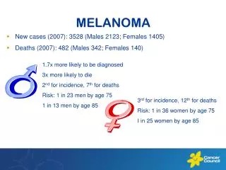
Melanoma. Kari Kendra MD, PhD 9/18/2009. Melanoma Incidence and Mortality. Incidence (US) 59,580 new cases 33,580 new male cases 26,000 new female cases 12 per 100,000 population Mortality (US) 7,770 total 4,910 males 2,860 females.


Melanoma
E N D
Presentation Transcript
Melanoma Kari Kendra MD, PhD 9/18/2009
Melanoma Incidence and Mortality • Incidence (US) • 59,580 new cases • 33,580 new male cases • 26,000 new female cases • 12 per 100,000 population • Mortality (US) • 7,770 total • 4,910 males • 2,860 females American Cancer Society,Cancer Facts and Figures. 2005.
Melanoma: risk factors • Constitutional predisposition • Fair skin/hair color/ freckling • Burn vs tan • >20 benign nevi (moles) or >3 atypical nevi • Family history of dysplastic nevi • Increasing age • Immunosuppression • Xeroderma pigmentosum • H/O solar keratosis, squamous cell carcinoma
Melanoma: risk factors • Risk behaviors • >3 sunburns • Episodic excessive sunlight exposure • Long term continuous sunlight exposure • UV exposure at tanning salons
Fifteen-year survival curves comparing localized melanoma (stages II and I), regional metastases (stage III), and distant metastases (stage IV) Balch, C. M. et al. J Clin Oncol; 19:3635-3648 2001
Melanoma The challenge (historically): • Early detection • Rapid growth/high proliferation rate • Chemotherapy resistant • Radiation resistant • Short anticipated survival
Asymmetry Border Benign Malignant Malignant
Color Diameter Benign Malignant Malignant
Prognostic indicators • Thickness (Breslow depth) • Nodal status • Ulceration • Mitosis • Satellite lesions • In transit lesions
Prognostic indicators • Thickness (Breslow depth) • Nodal status • Ulceration • Mitosis • Satellite lesions • In transit lesions
Prognostic indicators: Breslow depth Depth (mm)5-year survival (%) <0.75 96 0.76 – 1.49 87 1.50 – 2.49 75 2.50 – 3.99 66 >4.00 47 Koh HK, et al 1991
Biopsy techniques • Excisional biopsy 1-3 mm margins avoid wider margins (accurate lymphatic mapping) • Full thickness incisional/punch biopsy for large lesions lesions of the palms, soles, digits, face, ears • Deep shave biopsies When suspicion for melanoma is low NCCN Guidelines 2005
Surgical Treatment of Melanoma • In Situ: 0.5 mm margin wide excision • < 1.0 mm: 1 cm margin wide excision • 1.0 - 4.0 mm: 1-2 cm margin wide excision • > 4.0 mm: 2 cm margin wide excision
Prognostic indicators • Thickness (Breslow depth) • Nodal status • Ulceration • Mitosis • Satellite lesions • In transit lesions
Nodal evaluation • Tumors with depth >1 mm • Sentinel node evaluation • Tumors with depth < 1 mm • No nodal evaluation recommended NCCN Guidelines 2005
Negative Sentinel Lymph Nodes • 5 year survival >80% for Clarks level IV primaries • OS is 89% • Of the 11% who failed • 2.7% in the draining basin • 7.3% distant disease • Essner et al 2001
Fifteen-year survival curves for the stage groupings of patients with localized melanoma Balch, C. M. et al. J Clin Oncol; 19:3635-3648 2001
Prognostic Indicators: Nodal status • OS for patients with 1 positive sentinel node is 60% at 5 years • OS for patients with a single palpable node is 40% at 5 years • Gershenwald et al, 2001
Negative Sentinel Lymph Nodes Key prognostic indicators - ulceration and tumor thickness • T1 lesions (<0.75 mm) - 86% alive at 10 years • T4 lesions with ulceration (>4mm) – 10 year survival 41%
Prognostic indicators • Thickness (Breslow depth) • Nodal status • Ulceration • Mitosis • Satellite lesions • In transit lesions
Photomicrograph of a typical ulcerated melanoma Balch, C. M. et al. J Clin Oncol; 19:3635-3648 2001
Prognostic indicators • Thickness (Breslow depth) • Nodal status • Ulceration • Mitosis • Satellite lesions • In transit lesions
Mitotic Index • N = 3661 from the Sydney Melanoma Database • Correlated • clinical information (survival) • primary tumor thickness (Breslow depth) • ulcerative state (infiltrative, attenuative, and traumatic) • tumor mitotic rate (TMR) (at the invading front, deep border) • Conclusion: TMR is a more powerful prognostic indicator than ulceration in patients with primary cutaneous melanoma Azzola et al, Cancer 2003
Survival curves of 3661 patients with localized, cutaneous melanoma when tumor mitoses/mm2 were grouped into four categories .Azzola et al Cancer 2005 Azzola et al, 2003
Tumor thickness and Mitotic Index • Clarks level vs Mitotic Index • Clark level of invasion failed to have a significant prognostic impact by multivariate analysis after adjusting for TMR. • The number of patients with a Clarks level IV was too small for adequate comparison. (Azzola et al: Cancer 2005)
Prognostic indicators • Thickness (Breslow depth) • Nodal status • Ulceration • Mitosis • Satellite lesions • In transit lesions
Risk of In-Transit Metastasis • In- transit metastasis • Cutaneous / subcutaneous tissue • Between the primary tumor and the draining lymph node basin • 5 yr survival rates: 12% - 37% • Risk factors: • Thicker primary • Lower extremity • Regional LN metastasis
Other prognostic factors: • LDH • Elevated levels correlate with: Early recurrence Shorter survival (Newcki et al, 2008) • Serum S100 level • Early studies suggest: Shorter survival Early distant relapse Poorer response to treatment (Smith et al, 2008) • Microvessel Density
Other prognostic factors: • LDH • Elevated levels correlate with: Early recurrence Shorter survival (Newcki et al, 2008) • Serum S100 level • Early studies suggest: Shorter survival Early distant relapse Poorer response to treatment (Smith et al, 2008) • Microvessel Density
Other prognostic factors: • LDH • Elevated levels correlate with: Early recurrence Shorter survival (Newcki et al, 2008) • Serum S100 level • Early studies suggest: Shorter survival Early distant relapse Poorer response to treatment (Smith et al, 2008) • Microvessel Density
Microvessel Density (MVD) • N= 244 • Histology: superficial spreading (59.3%), nodular (21.1%), lentigo maligna melanoma (9.8%), acral lentiginous (4.4%) • Breslow thickness: 0.1 mm – 14.8 mm • Duration of follow-up : minimum of 2 years • Immunostain: with CD31 antibody to identify endothelium • Vessel count performed at the tumor edge and in the peritumoral host tissue and in the tumor center for depth >2mm Depasquale et al Histopathology 2005
Microvessel Density (MVD) • Conclusion: microvessel assessment of primary melanoma using the Chalkley score technique provides reliable prognostic information on the risk of recurrence of the tumor, particularly for melanomas deeper than 2 mm. • Chalkley score – total number of microvessels and relative area occupied by vasculature Depasquale et al Histopathology 2005
Melanoma The challenge (historically): • Early detection • Rapidly growing • Chemotherapy resistant • Radiation resistant • Short anticipated survival
Fifteen-year survival curves for the stage groupings of patients with localized melanoma Balch, C. M. et al. J Clin Oncol; 19:3635-3648 2001
Adjuvant therapy for high risk patients What therapies are available? How do we identify patients for treatment?
Case 1 34 y/o female presented with a bleeding mole on her arm. • Biopsy: nodular melanoma, 4.1 mm deep, with ulceration, mitotic rate 15/10 HPF • Wide excision: no residual tumor • Sentinel Node: positive for 2/2 LN • Axillary LN dissection: 0/20 LN
Case 1 What is the next step?
Systemic Therapy:Adjuvant • Biologic Agents • IL2 • IFN • GM-CSF • Chemotherapeutic agents • Cisplatin • Vinblastine • DTIC • Biochemotherapy
Adjuvant therapy with Interferon Alfa-2b (E1684) FDA approved • IFN-alpha 2b for adjuvant treatment of melanoma patients with thick primary tumors (> 4mm) or resected nodal disease • positive response data is for node + patients only
Adjuvant therapy with Interferon Alfa-2b (E1684) • Patient population • Breslows depth >4mm • LN+ after ELND • clinical LN+ with synchronous primary • regional LN recurrence after surgery for primary Kirkwood et al, JCO 1996;14:7
Adjuvant therapy with Interferon Alfa-2b (E1684) Treatment • high-dose IFNα-2b : 20 MU/m2 IV, 5 days per week for 4 weeks (induction phase) followed by 10 MU/m2 SC TIW for 48 weeks • observation
Adjuvant therapy with Interferon Alfa-2b (E1684) IFN-2bObservation median DFS 1.7 yr 1.0 yr OS 3.8 yr 2.8 yr • benefit greatest in LN+ patients • benefit most pronounced early during the treatment interval
Adjuvant therapy with Interferon Alfa-2b (E1684) TOXICITIES: constitutional myelosuppression hepatotoxocity neurologic • 67% of all patients had severe (grade 3) toxicity at some point during treatment
Adjuvant therapy with Interferon Alfa-2b (E1684) Time from patients receiving 80% of randomizationtarget dose induction 67% 3 months 62% 6 months 40% 9 months 30% 11 months 25%