CEREBELLAR DYSFUNCTIONS
CEREBELLAR DYSFUNCTIONS LECTURE-30 (20 APRIL 2013) 1-2 PM Professor Dr Wahengbam PS AL Waheed MBBS,MD(PSM),MD(Medicine ).

CEREBELLAR DYSFUNCTIONS
E N D
Presentation Transcript
CEREBELLAR DYSFUNCTIONS LECTURE-30 (20 APRIL 2013) 1-2 PMProfessor DrWahengbam PS AL WaheedMBBS,MD(PSM),MD(Medicine)
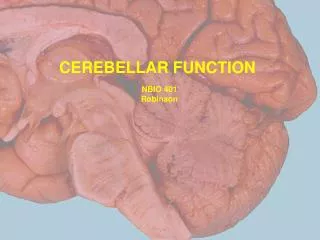
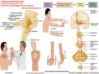
Recall cerebellar functionsThe cerebellum processes input from other areas of the brain, spinal cord and sensory receptors to provide precise timing for coordinated, smooth movements of the skeletal muscular system. The cerebellum can be divided into central structures lingula, vermis and flocculonodular lobe and the cerebellar hemispheres. The cerebellum has 3 parts: Archicerebellum (vestibulo cerebellum): It includes the flocculonodular lobe, which is located in the medial zone. The archi cerebellum helps maintain equilibrium and coordinate eye, head, and neck movements; it is closely interconnected with the vestibular nuclei.Midline vermis (paleo cerebellum): It helps coordinate trunk and leg movements. Vermislesions result in abnormalities of stance and gait.Lateral hemispheres (neocerebellum): They control quick and finely coordinated limb movements, predominantly of the arms.Main inputs come from frontoponto cerebellar connections (contralateral) from above, and spino cerebellar tracts from below (proprioception) producing primarily ipsilateral signs. In addition to coordination, the cerebellum controls some aspects of memory, learning, and cognition.Ataxia is the archetypal sign of cerebellar dysfunction, but many other motor abnormalities may occur
Function The strongest clues to the function of the cerebellum have come from examining the consequences of damage to it. Humans cerebellar dysfunction show problems with motor control, on the side of the body ipsilateral to the damaged cerebellum. They continue to be able to generate motor activity, but it loses precision, producing erratic, uncoordinated, or incorrectly timed movements. A standard test of cerebellar function is to reach with the tip of the finger for a target at arm's length: A healthy person will move the fingertip in a rapid straight trajectory, whereas a person with cerebellar damage will reach slowly and erratically, with many mid-course corrections. PrinciplesFour principles have been identified as important: 1. Feed forward processing: The signals move unidirectionthrough the system from input to output, with very little recurrent internal transmission. This feed forward mode of operation means that Signals enter the circuit, are processed by each stage in sequential order, and then leave. 2. Divergence and convergence: Information from 200 million mossy fiber inputs is expanded to 40 billion granule cells, whose parallel fiber outputs then converge onto 15 million Purkinje cells. Thus, the cerebellar network receives a modest number of inputs, processes them very extensively through its rigorously structured internal network, and sends out the results via a very limited number of output cells.
3. Modularity: A module consists of a small cluster of neurons in the inferior olivary nucleus, a set of long narrow strips of Purkinje cells in the cerebellar cortex (microzones), and a small cluster of neurons in one of the deep cerebellar nuclei. Different modules share input from mossy fibers and parallel fibers, but appear to function independently — the output of one module does not appear to significantly influence the activity of other modules.4. Plasticity: The synapses between parallel fibers and Purkinje cells, and the synapses between mossy fibers and deep nuclear cells, This arrangement gives tremendous flexibility for fine-tuning the relationship between cerebellar inputs and outputs.One of the most extensively studied cerebellar learning tasks is the eye blink conditioning paradigm, in which a neutral conditioned stimulus such as a tone or a light is repeatedly paired with an unconditioned stimulus, such as an air puff, that elicits a blink response.
A stroke affecting the cerebellum may cause dizziness, nausea, balance and coordination problems.Congenital malformations: often occurring as part of complex malformation syndromes --Congenital Neurologic Manifestations vary markedly depending on the structures involved BUT, ataxia is usually present.Hereditary ataxias:results from a gene mutation causing abnormal repetition of the DNA sequence. Gait unsteadiness, dysarthria, and paresis, Mental function often declines. Tremor, is slight. Reflexes and vibration and position senses are lost. Talipesequinovarus (clubfoot), scoliosis, and progressive cardiomyopathy are common. By their late 20s, patients may be confined to a wheelchair. Death, often due to arrhythmia or heart failure, usually occurs by middle age.Spinocerebellarataxias autosomal dominant ataxias. Manifestations are multiple areas in the central and peripheral nervous systems; neuropathy, pyramidal signs, and restless leg syndrome, ataxia, are common. Symptoms include ataxia, parkinsonism, and possibly dystonia, facial twitching, ophthalmoplegia, and peculiar bulging eyes.Drug induced cerebellar dysfunction and ataxiaPhenytoin, and Toxic levels of certain drugs (Anticonvulsants) In children, primary brain tumors (medulloblastoma, cystic astrocytoma) in the midline cerebellum and reversible diffuse cerebellar dysfunction f due to viral infections.,
Identify cerebellar dysfunctionCerebellar ataxia Lesions of the midline vermis of the cerebellum cause truncal ataxia, while lesions of the cerebellar hemispheres cause limb ataxia of the ipsilateral side.Gait ataxiaPatientswill tend to stand with feet well apart and are often frightened to stand. Patients tend to reel to the side of unilateral lesion, or from side to side if central or bilateral (even if supported). Walking along a line of the floor demonstrates minor degrees of gait ataxia. Wobbling may increase if eyes are closed but patients don't fall - this is not a true "positive Romberg's test" (which is positive when there is impaired joint proprioception).TruncalataxiaPatientscan't sit or stand unsupported and tend to fall backwards. It is caused by a midline cerebellar lesion, or may be a feature of post-chickenpox cerebellar syndrome. Truncal tremor may be evident - constant jerking of trunk and head.Limb ataxiaLesionsof the cerebellar hemisphere cause ipsilateral signs. The outstretched arm tends to be held hyperpronated at rest and at a slightly higher level than unaffected side (Riddoch's sign), and rebounds upwards if gently pressed downwards and then suddenly released by the examiner.
Finger-nose and heel-knee-shin tests will demonstrate even mild limb ataxia, with terminal intention tremor and dysmetria (past pointing).Cerebellar dysarthria Aspluttering staccato speech. Scanning dysarthria - jerky and explosive speech with separated syllables may be demonstrated by asking the patient to repeat "baby hippopotamus".WritingThismay be larger than normal (contrast with micrographia of Parkinson's disease).Rapid alternating movementsCerebellarlesions produce inaccuracies in rapidly repeated movements (dysdiadochokinesia). This is demonstrated by getting the patient to tap the back of their own hand repeatedly with the other hand, or to tap their foot on the floor.TremorUnilateralor bilateral intention tremor, or a truncal tremor.Nausea and vomitingSudden vomiting (without warning) after a positional change, without preceding nausea, is suggestive of a posterior fossa lesion.
Vascular: Stroke or transient ischemic attack (TIA)Infarction of the posterior inferior cerebellar artery causes lateral medullary syndrome with hemiataxia, vertigo, dysarthria, ptosis and miosisSpace-occupying HydrocephalusEnlarging masses in the cerebellum may obstruct CSF flow, causing hydrocephalus and raised intracranial pressure. Posterior fossa tumours or abscess.Coning of the cerebellar tonsils can occur rapidly (within hours), causing respiratory arrest.Nutritional:Thiamine deficiency - Wernicke's encephalopathy (triad of acute confusion, ataxia and ophthalmoplegia); Vitamin E deficiency Gluten sensitivity (gluten ataxia):
Infections:Bacterial: meningo-encephalitis or intracranial abscessViral: acute infections (varicella); chronic infections, HIV; post-viral syndromes (in childhood)Parasitic infections (toxoplasma, falciparum malaria, Lyme disease)Prions: Creutzfeldt-Jakob disease Toxins: alcohol, mercury, other heavy metals, solvents, carbon monoxide poisoningDrugs: barbiturates, phenytoin, piperazine, antineoplastic drugs, deferiproneTraumaMultiple sclerosis (MS)Genetic: Friedreich'sataxia and ataxia telangiectasiaMetabolic and endocrine: Cerebral oedema of chronic hypoxia Wilson's disease , HypothyroidismCongenital:Cerebral palsyIdiopathic cerebellar ataxia
Review clinical cases related to cerebellar dysfunctionAs the cerebellum is associated with motor control, lesions produce a range of movement disorders (ataxias). Acute onset ataxiaEitherdue to cerebellar haemorrhage or infarction. Haemorrhagepresents with:Occipital headacheVertigoVomitingAltered consciousnessSubacute ataxiaMay occur from: Viral infection - present with pyrexia, limb and gait ataxia, dysarthria appearing over hours or days; takes up to six months for full recoveryPost-infectious encephalomyelitis - commonly related to varicella Other causes include - hydrocephalus, posterior fossa tumours, abscesses, parasitic infections and various toxinsEpisodic ataxias This is episodes of ataxia lasting minutes to hours. Due to Drugs MS Transient vertebrobasilarischaemic attacks Foramen magnum compression
Review clinical cases related to cerebellar dysfunction( contd.)Chronic progressive ataxias Commonly caused by chronic alcohol abuse with malnutrition may improve with thiamineIngestion of drugs - Anticonvulsants, Phenytoin, Heavy metalsStructural lesionsParaneoplasticcerebellar degeneration Carcinomas of the lung or ovariesCerebellar disorders in infantsOther causesPontocerebellar hypoplasia Joubert'ssyndromeTrisomiesPyruvate dehydrogenase deficiencySpastic ataxic syndrome
Midline lesions can produce severe gait and truncal ataxia. As they extend they can also give fourth cranial nerve lesions and severe ipsilateral arm tremor, marked nystagmus, vertigo and vomiting, and can block CSF flow (obstructive hydrocephalus). Cerebellar hemisphere lesions can produce classic ipsilateral limb ataxia (intention tremor, past pointing and mild hypotonia). Limb rebound can be demonstrated by gently pushing down on outstretched arms and then suddenly releasing, causing the arm on the affected side to suddenly fly upwards. Lateral lesions tend to produce more subtle nystagmus (maximal looking towards side of lesion). Ataxiais the archetypal sign of cerebellar dysfunction, but many other motor abnormalities may occur
Check eye movement - looking for ophthalmoplegia or nystagmusFundi for papilloedemaTongue out and move it from side to side (movement slowed)Ask patient to repeat "baby hippopotamus" - look for dysarthria and abnormal speechExamine arms for limb ataxia : rebound of outstretched arms, finger-nose test for past pointing, check for dysdiadochokinesis,heel-shin test,Mild hypotonia and hyporeflexia,Sit up with arms crossed - looking for truncalataxia,Walk heel-to-toe (to elicit any gait ataxia)Stand with feet together - unsteady with eyes open and closed . ( Romberg's positive)
InvestigationsThese should be guided according to the differential diagnosis based upon the initial assessment. This may include:Blood tests – full blood count, liver function tests, cholesterol, protein electrophoresis, copper and caeruloplasmin, immunoglobulins and glycoproteinsEEGEMGImaging - MRI is the modality of choiceManagement and prognosisThis depends upon the underlying cause.