Protein Tyrosine Phosphatases (PTPs)
570 likes | 1k Vues
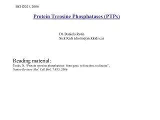
BCH2021, 2006. Protein Tyrosine Phosphatases (PTPs). Dr. Daniela Rotin Sick Kids (drotin@sickkids.ca). Reading material: Tonks, N, “Protein tyrosine phosphatases: from gene, to function, to disease”, Nature Reviews Mol. Cell Biol . 7:833, 2006. Lectures overview : 1. PTPs :

Protein Tyrosine Phosphatases (PTPs)
E N D
Presentation Transcript
BCH2021, 2006 Protein Tyrosine Phosphatases (PTPs) Dr. Daniela Rotin Sick Kids (drotin@sickkids.ca) Reading material: Tonks, N, “Protein tyrosine phosphatases: from gene, to function, to disease”, Nature Reviews Mol. Cell Biol. 7:833, 2006
Lectures overview: 1. PTPs: General overview Activity Classification Dimerization Oxidation 2. Receptor PTPs: LAR family CD45 PTPalpha/epsilon 3. Non-receptor PTPs: PTP.1B SHP-1, SHP-2 4. Non-classic PTPs PTEN
Tyrosine phosphorylation and dephosphporylation Y In mammals: ~90 Tyr kinases ~107 Tyr Phosphatases (PTPs)
Protein Tyrosine Phosphatases (PTPs) 1. Although originally thought of as constitutively active enzyme that dephosphorylate Tyr phosphorylated proteins (opposing tyrosine kinase activity), it appears that PTPs are highly regulated, specific and are equally important as Tyr kinases. 2. Because Tyr kinases are regarded as promoting cell signaling and proliferation, it was initially assumed that PTPs would inhibit signaling and cell proliferation. This is not true: Some PTPs have inhibitory and some stimulatory roles in regulating cell proliferation. 3. PTPs are NOT related to Ser or Thr phosphatases
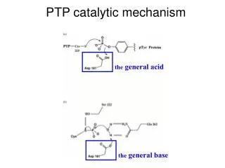
Due to the dependence on the catalytic Cys in this reaction, PTPs are very sensitive to oxidation state of the cell (they are inhibited by oxidation)
Tools to identify substrates for PTPs: “Substrate Trapping” PTPs are rendered catalytically inactive by mutating the Cys (C215 in PTP.1B) or Asp (D181 in PTP.1B) needed for catalysis. They still allow binding to substrates but not dephsophorylating them, hence the substrates become “Trapped” (bound) and can then be identified.
Classic PTPs CLASSIFICATION: (R=Receptor; NR=Non Receptor) Cell Adhesion molecule
Other PTPs (DSP=Dual Specificity Phosphatase)
Substrate specificity
Dimerization of Receptor PTPs (compared to receptor Tyrosine kinase, RTK)
X X Interactions between Receptor PTPs lead to inhibition of catalytic activity: Intra- or inter- molecular interactions between the first and second phosphatase domain. Eg 1) In LAR family PTPsD2 (catalytically-inactive) binds D1, and inhibits its catalytic activity 2) In PTPalpha, CD45: The first catalytic domain (D1) binds the Wedge region of another D1 domain (homo-dimerization) LAR family PTPs Ig(x3) The inactive second catalytic domain of LAR family PTPs (eg PTPdelta) binds to and inhibits the activity of the first catalytic domain (of eg PTPsigma). FNIII (x8) TM D1 D2 PTPalpha The first catalytic domain of PTPalpha binds to the wedge region and inhibits the activity of the first catalytic domain of another PTPalpha (both first and second catalytic domains are active) TM Wedge D1 D2
H2O2 Oxidation of PTP active site: H2O2 incubation with the catalytic domain of PTP.1B leads to conformational changes. The PTP loop (containing the signature motif) and the Tyr from the pTyr binding loop, which are normally buried, flip out of the active site and become solvent-exposed. Does this occur in vivo?
Regulation of RPTP function by dimerization: a new role for oxidation H2O2 Oxidation leads to stabilization of the inactive state of RPTPs promoting S-S bonds of the Cys in the second catalytic domain (D2)
Receptor PTPs LAR family PTPs: Role in development (especially the nervous system)
Signaling downstream of LAR family PTPs Role of the extracellular domain: -A cell adhesion molecule -PTPdelta and LAR: Homophilic interactions of ectodomain promotes neurite growth and axon guidance -LAR ectodomain( missing FN5 repeat) binds laminin-nidogen complex (but biological consequence of this binding is not known) -PTPsigma: Heterophilic interactions: ectodomain binds heparan sulphate proteoglycan (HSPG) (agrin and collagen XIII) in chick retinal axons. In muscle, different ligands are likely important (not HSPG). This binding does not affect catalytic activity. Ie. Biological function of ligand binding not completely understood. Role of Intracellular domain: -Generally linked to remodeling of the actin cytoskeleton -Liprins are LAR family interacting proteins, but are not substrates. Rather, they serve as scaffold proteins important for maintenance of the presynaptic zone, and they bind GRIP and GIT (glutamate receptor interacting proteins). They are also important at the post-synaptic zone. -Trio binds LAR. Trio is a guanine nucleotide exchange factor for Rac1 and RhoA, which are important for cytoskeleal remodeling. Trio also binds Focal adhesion kinase (FAK) and is phosphorylated by FAK. Trio also binds MIM-B, a binding partner to PTPdelta. -The tyrosine kinase Abl and its substrate Ena are involved in axon growth and guidance, and oppose function of DLAR in Drosophila (flies). LAR likely dephosphorylates Ena (ie Ena is its substrate). -LAR and PTPsigma bind to and dephosphorylates N-cadherin and beta-catenin. The catenin complex is itself in complex with actin. ie it connects between cadherin and the actin cytoskeleton, and is important for axon growth. *Additional roles: -LAR binds the Insulin receptor and dephosphoryaltes it in vitro, although In vivo this may be a result of dephosphorylation of the IR substrates IRS-1 and -2. Hence LAR regulates insulin signaling.
Biological functions of LAR family PTPs: • Mainly studies in the nervous system, and examples from flies and mammals are provided in • the following slides
LAR family PTPs regulate muscle synaptogenesis in embryonic flies
LAR family PTPs regulate axon guidance in the developing retina of flies
Role of LAR family PTPs in mammalian development: LAR family members are strongly expressed in the developing nervous system as well as in several Epithelial tissues (eg. lung, intestine) 1.Neuroendocrine:PTPsigma regulates Growth Hormone and Prolactin secretion from the pituitary 2. Neuronal: PTPsigma attenuates neurite outgrowth and nerve regeneration (see next slide), while LAR accelerates them. PTPsigma also regulates axon guidance. PTPdelta also involved in regulating CNS function (learning and memory) 3. Pancreas: LAR (and probably PTPsigma): involved in regulation of insulin signaling downstream of the Insulin Receptor and hence blood glucose levels (reveal insulin resistance). LAR family and cancer: Not much is known, and mostly correlative data (eg increase LAR expression in metastatic breast cancer, and PTPsigma reduces colony formation (indicative of cancer) in soft agar assay)
CD45: Role in regulation of the immune system
The Tyr kinase LCK is required for T cell activation. LCK is inactive at resting state (its SH2 domain binds its own Tyr phosphorylated Y505 and the SH3 domains binds a PPII loop). CD45 dephosphorylates the LCK-Y505 site, thus relieving the inhibition and allowing LCK to become active by phosphorylating Tyr 394. • Ie CD45 has a POSITIVE role in T cell activation PPII • CD45 deficient mice, or patients with mutations in CD45, suffer from severe combined immune deficiency • CD45 is a positive regulator of antigen (in T cells) and Ig receptor (in B cells) signaling. In T cells it dephosphorylates • LCK, FYN and YES Tyr kinases, and in B cells it dephosphorylates LYN, FYN and BLK
Regulation of CD45: Dimerization -Homo-dimerization of CD45 inhibits its catalytic activity, thus attenuating T cell activation. (mutation in the Wedge sequence of CD45 in mice relieves its inhibition of phosphatase activity, leading to enhanced CD45 and TCR activation, and to lymphoproliferative disease and autoimmunity) -The tendency to dimerize is greater with the CD45RO isoform (which is less glycosylated). 2) Spatio-temporal: -CD45 association with lipid rafts (where the TCR complex is found) is important for its regulation of TCR activation. 3) Phosphorylation CD45 is phsophorylated (by CK2, PKC, CSK), but the physiological significance of this is unknown.
PTPalpha, PTPepsilon Regulation of Src signaling and cancer
PTPalpha (and PTPepsilon) activate Src by dephosphorylating its inhibitory site (Tyr527). This leads to enhanced cell proliferation (ie PTPalpha has a positive role in regulating cell signaling). It may also have a role in cancer (reminiscent of v-Src which is constitutively active). v-Src (viral Src) (Oncogenic) c-Src SH3 SH3 SH2 SH2 SH1(catalytic) SH1(catalytic) Y527 Dephosphorylated (activated) by PTPalpha
Biological functions of PTPalpha and PTPepsilon: Activates Src (or other Src family kinases), leading to increased cell proliferation and transformation Important for integrin-mediated signaling and hence cell adhesion/spreading Important for hippocampal neuronal migration and long term potentiation Regulates K channels and the NMDA receptors Important for bone homeostasis (PTPepsilon)
Non Receptor-PTPs PTP.1B: Role in regulation of metabolism
PTP.1B • Earlier work had demonstrated that the PTP inhibitor Vanadate increased insulin signaling • PTP.1B is a regulator of insulin action and binds the activated Insulin receptor (IR) (see next slide) • PTP.1B becomes Tyr phosphorylated following binding to the IR • PTP.1B dephosphorylates the IR (and possibly IRS-1, the IR substrate), hence it is a negative • regulator if insulin signaling. Ie it regulates cellular metabolism, not mitogenesis. • PTP.1B knockout in mice leads to defects in glucose and insulin tolerance • (increased insulin sensitivity) in muscle and liver (but not adipose tissues) • PTP.1B was shown to regulate obesity by regulating Leptin’s action • PTP.1B is widely expressed. Localized to the ER, but a cleavable C terminus allows it • to enter the cytosol. • Because PTP.1B knockout mice show increased insulin sensitivity and quick clearance of glucose • from the blood, extensive search is now being carried out to identify inhibitors of PTP.1B, in order • to treat type II diabetes and obesity. • Knockdown of PTP.1B (with antisense oligonucleotides) in obese mice (ob/ob) resulted in • alleviation of insulin resistance • PTP.1B knockout mice are resistant to diet-induced obesity(and show enhanced leptin sensitivity). • But, a role of PTP.1B in leptin signaling is not yet clear.
SHP-1, SHP-2: (SH2- domain containing PTPs) Role in immune regulation, signaling and human diseases Diseases associated with mutations in SHP-1 and SHP-2: SHP-1: autoimmune disease SHP-2: Noonan Syndrome (NS), Leukemias, Leopard Syndrome (NS: short stature, characteristic facial features, heart defects. Incidence: 1:2000 live birth. 50% of patients have mutations in SHP-2) LS: lentigines (sun spots), ECG abnormalities, Pulmonic valve stenosis, deafness, growth retardation)
SHP-1 (Primarily expressed in hematopoietic cells) SH2 SH2 PTP • - Motheaten mice have a frameshift mutation that truncates most of the Shp-1 gene • (they are thus considered null). • They die of auto-immune disease primarily affecting their myeloid cells • SHP-1 provides a negative regulatory signals in the immune system • (as oppose to SHP-2, which provides positive signal for cell proliferation) • SHP-1 negatively regulates integrin signaling, TNF receptor signaling and EpoR signaling
SHP-2 S=substrate BP=binding protein
SHP-2 SHP-2 is widely expressed in many tissues. SHP-2 deficiency: -In flies (Drosophila): SHP-2(CSW) affects signaling downstream of tyrosine kinase growth factor receptors (eg EGFR, FGFR). SHP-2/CSW plays a positive (signaling enhancing) role, hence its deficiency affects normal development -In frogs (Xenopus): SHP-2 is required for gastrulation and FGFR-dependent development -In mice: SHP-2 null mice are embryonic-lethal (ie SHP-2 is needed for mammalian development likely downstream of the FGFR)
Regulation of SHP-2 activity c. NS=Noonan syndrome JMML=Juvenile myelomonocytic Leukemia
Noonan Syndrome/Leukemia: Gain of function mutations Leopard syndrome: Dominant negative (catalytically-inactive) mutations
SHP-2 signaling: possible models b) SHP-2 is recruited by the adaptor Gab1 to dephsophorylate the inhibitory Tyr on Src (Tyr 527), activating Src, which in turn activates the Ras/Erk pathway. Alternatively, SHP-2 may dephosphorylate and inactivate CSK, the Tyr kinase that phosphorylates (and inactivates) Src-Y527. a) SHP-2 dephosphorylates the RasGAP binding site on the receptor, thus blocking rasGAP and enhancing Ras/Erk signaling c) SHP-2 may dephosphorylate and inactivate Sprouty, the Ras signaling inhibitor (which must be phosphorylated in order to inhibit Ras). Another possibility is the SHP-2 interferes with signaling downstream of Erk
PTEN PDZ binding motif C2 Phosphatase 1. Although PTEN has the PTP signature motif in its phosphatase domain, it actually dephosphorylates Phospholipids 2. PTEN is a tumor suppressor