Download
1 / 62
620 likes | 864 Vues
Arrhythmia 341. Ahmad Hersi , MBBS, MSc,FRCPC Asso . Professor of Medicine& Consultant Electrophysiologist. Objectives. Identify mechanism of AF Recognize EKG of AF Discuss treatment options of AF Identify other forms of Arrhythmia.

E N D
Arrhythmia 341 Ahmad Hersi, MBBS, MSc,FRCPC Asso. Professor of Medicine& Consultant Electrophysiologist
Objectives • Identify mechanism of AF • Recognize EKG of AF • Discuss treatment options of AF • Identify other forms of Arrhythmia
Atrial fibrillation accounts for 1/3 of all patient discharges with arrhythmia as principal diagnosis. • 6% PSVT • 6% PVCs • 18% Unspecified 2% VF • 4% Atrial Flutter • 9% SSS • 34% Atrial Fibrillation • 8% Conduction Disease • 10% VT • 3% SCD
Epidemiology • 2.3 million people in North America • 4.5 million in EU • In the 20 year AF admission have increased by 66%. • $ 15.7 billion annually in EU • Estimated prevalence of AF is 0.4% to 1% in the general pop. 8% in pt. >80 years
Pathophysiology of atrial fibrillation and associated stroke
Normal regulation of heart rate and rhythm • Contraction is controlled by the sinoatrial (SA) node
Normal heart rhythm is disrupted in AF • AF is characterized by: • Rapid (350–600 beats/min) and irregular atrial rhythm • Reduced filling of the left and right ventricles • Conduction of most impulses from the atria to ventricles is blocked at the AV node • Contraction of the ventricles can be: • Irregular and rapid (110–180 beats/min; tachycardia) • Irregular and slow (<50 beats/min; bradycardia) • Normal • Cardiac output can be reduced
AF begets AF • AF causes remodeling that contributes to the initiation and maintenance of AF, including: • Electrical: shortening of refractory period • Structural: enlargement of atrial cavities • Initially, many episodes of AF resolve spontaneously • However, over time AF tends to become persistent or permanent due to electrical and structural remodeling Wijffels MC et al. Circulation 195;92:1954–68
Consequences of AF • Formation of blood clots (thrombosis) on the walls of the atria that can dislodge (embolize), leading to stroke and systemic embolism • Reduction in cardiac output can precipitate heart failure leading to: • Peripheral oedema • Pulmonary oedema
Diagnosis of AF • Signs and symptoms • Electrocardiography • Transthoracic echocardiography • Laboratory tests • Holter monitoring • Transoesophageal echocardiography • Exercise testing • Chest radiography
Heterogeneous clinical presentation of AF • With or without detectable heart disease • Episodic • Symptoms may be absent or intermittent • Up to 90% of episodes may not cause symptoms • Symptoms vary according to • Irregularity and rate of ventricular response • Functional status • AF duration • Patient factors • Co-morbidities Fuster V et al. Circulation 2006;114:e257–e354; Page RL et al. Circulation 1994;89:224–7
Signs and symptoms Fuster V et al. Circulation 2006;114:e257–354
All patients History Physical examination Electrocardiogram (ECG) Transthoracic echocardiogram (TTE) Blood tests Holter monitor Chest x-ray Selected patients Transesophageal echocardiogram (TEE) Clinical evaluation of patients with AF Adapted from Fuster V et al. Circulation 2006;114:e257–354
History and physical examination • Clinical conditions associated with AF • Underlying heart conditions (e.g. valvular heart disease, heart failure, coronary artery disease, hypertension) • Other reversible conditions • Family history • Familial AF (lone AF in a family) • AF secondary to other genetic conditions(familial cardiomyopathies) • Type of AF • First episode, paroxysmal, persistent, permanent • Triggers – e.g. emotional stress, alcohol, physical exercise, gastroesophageal disease • Specific symptoms • Response to any treatments administered Fuster V et al. Circulation 2006;114:e257–354;de Vos CB et al. Eur Heart J 2008;29:632–9
Electrocardiogram • Assesses the electrical activity of the heart • Essential for all patients with suspected AF, to identify • Abnormal heart rhythm (verify AF) • Left ventricular hypertrophy • Pre-excitation • Bundle-branch block • Prior MI • Differential diagnosis of other atrial arrhythmias Fuster V et al. Circulation 2006;114:e257–354
Eletrocardiogram: normal sinus rhythm • Impulse from sinoatrial (SA) node stimulates myocardiumto contract • P-wave:atrial depolarization • QRS complex:ventricular depolarization • T-wave: ventricular repolarization
P No P Electrocardiogram: loss of P wave in AF • Normal sinus rhythm • Normal heart rate • Regular rhythm • P Waves • Steady baseline • AF • Heart rate increased (tachyarrhythmia)* • Irregular rhythm • No P wave • Irregular baseline • *Reduced heart rate (bradyarrythmia) may also be observed
Transthoracic echocardiography (TTE) • Non-invasive • Used to identify • Size and functioning of atria and ventricles • Ventricle hypertrophy • Pericardial disease • Valvular heart disease
Laboratory tests • Routine blood tests should be carried out at least oncein patients with AF • Important parameters to assess include: • Thyroid function • Renal function • Hepatic function • Serum electrolytes • Complete blood count
Holter monitor • Portable ECG device • Continuous monitoring for a short period of time (typically 24 hours) • Useful for • Detecting asymptomatic AF • Evaluating patients with paroxysmal AF • Associating symptoms with heart rhythm disturbance • Assessing response to treatment
Transoesophageal echocardiogram (TEE) • Ultrasound transducer positioned close to the heart using an endoscope-like device • High quality images of cardiac structure and function • Particularly the left atrial appendage, the most common site of thrombi in patients with AF • Not routinely used but useful for: • Accurate assessment of risk of stroke • Detection of low flow velocity (‘smoke’ effect) • Sensitive detection of atrial thrombi
Chest Radiography • When clinical findings suggest an abnormality chest radiography may be used to • Evaluate pulmonary pathology and vasculature • Detect congestive heart failure • Assess enlargement of the cardiac chambers
Classification of AF: joint guidelines of the ACC, AHA and ESC (1)
Classification of AF: joint guidelines of the ACC, AHA and ESC (2)
3 Strategies • Prevention of thromboembolism • Rate control • Restoration and maintenance of sinus rhythm
Treatment options for AF STROKE PREVENTION CONTROL OF HEART RATE MAINTENANCE OF SINUS RHYTHM PHARMACOLOGIC PHARMACOLOGIC PHARMACOLOGIC • Warfarin • Aspirin • Dabigatran • Ca2+-channel blockers • -blockers • Digoxin • Antiarrhythmic drugs • – Class IA • – Class IC– Class III: e.g. amiodarone, dronedarone NON-PHARMACOLOGIC NON-PHARMACOLOGIC NON-PHARMACOLOGIC • Removal/isolation of left atrial appendage, e.g. WATCHMAN® device or surgery • Ablate/pace • Ablation • Surgery (MAZE) ACE = angiotensin-converting enzyme 30
Rhythm-control therapies The objective of rhythm-control therapy is to restore (cardioversion) and maintain normal sinus rhythm Cardioversion can be achieved by: Pharmacotherapy with antiarrhythmic agents Electrical shocks (direct-current cardioversion) Direct-current cardioversion is generally more effective than pharmacotherapy Likelihood of successful cardioversion decreases with the duration of AF Pharmacological cardioversion is most effective when initiated within 7 days of AF onset Cardioversion can dislodge thrombi in the atria, increasing the risk of stroke Thromboprophylaxis is recommended for 3 wk before and for at least 4 wks after cardioversion in patients with AF that has persisted for 48 h 37 Fuster V et al. Circulation 2006; 114:e257–354
TEE-guided cardioversion: ACUTE study design AF >2 d durationDirect-current cardioversion prescribed TEE-guided cardioversion Conventional cardioversion Therapeutic anticoagulationwith heparin or warfarin Thrombus No thrombus No cardioversion Cardioversion Warfarin for 3 wk Warfarin for 3 wk Repeated TEE Warfarin for 4 wk Cardioversion No thrombus Thrombus Cardioversion No cardioversion Warfarin for 4 wk Warfarin for 4 wk Warfarin for 4 wk Follow-up (8 wk) DC = direct-current; TEE = transoesophageal echocardiography Klein AL et al. N Engl J Med 2001;344:1411–20
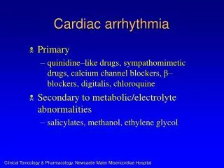
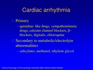
Atrial Fibrillation: Cardiac Causes • Hypertensive heart disease • Ischemic heart disease • Valvular heart disease • Rheumatic: mitral stenosis • Non-rheumatic: aortic stenosis, mitral regurgitation • Pericarditis • Cardiac tumors: atrialmyxoma • Sick sinus syndrome • Cardiomyopathy • Hypertrophic • Idiopathic dilated (? cause vs. effect) • Post-coronary bypass surgery
Atrial Fibrillation: Non-Cardiac Causes • Pulmonary • COPD • Pneumonia • Pulmonary embolism • Metabolic • Thyroid disease: hyperthyroidism • Electrolyte disorder • Toxic: alcohol (‘holiday heart’ syndrome)
Background - Atrial Flutter • Underlying mechanism – large “macro re-entrant circuit” in the atrium, typically moves counter-clockwise • Atrial rate range: 250-350 bpm • Ventricular response depends on the degree of AV block: • 2:1 block ventricular rate = 150 bpm • 3:1 block 100 bpm • 4:1 block 75 bpm
Causes – Atrial Flutter • Most commonly occurs in male patients with dilated or distended atria with elevated left atrial pressure • Clinical scenarios: • Systolic CHF with low EF • Mitral regurgitation (MR)
Rx – Atrial Flutter • Unstable pt (i.e. low BP / CP / AMS): • Synchronized cardioversion as per ACLS • 50J 100J 200J 300J 360J • Stable pt: • Rate control - just like atrial fibrillation (AFib) • Elective cardioversion - just like AFib • Anti-coagulation – just like AFib • Refer for Ablation
So What Is Actually Meant By Supraventricular Tachycardia? • Arrhythmias of supraventricular origin using a re-entrant mechanism with abrupt onset & termination • AVNRT (60%) • AVRT (30%) • Atrial tachycardia (10%)
3 Steps in Diagnosing Tachyarrhythmias • Step#1 - determine if the QRS complex is narrow or wide • Step #2 - determine if the QRS complexes are regular or irregular • Step #3 - look for P waves and their relationship to the QRS complexes
3 Categories of Tachyarrhythmias • #1 - Narrow complex / Regular rhythm • #2 - Narrow complex / Irregular rhythm • #3 - Wide complex
Atrioventricular Nodal Re-entrant Tachycardia (AVNRT) or AVRT