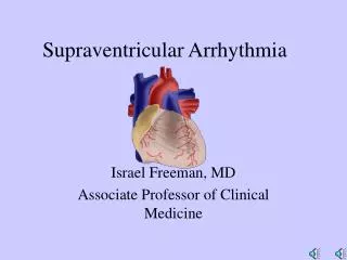
Arrhythmia
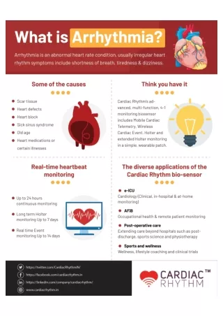
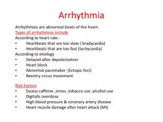
Arrhythmia. Arrhythmias are abnormal beats of the heart. Types of arrhythmias include : According to heart rate : Heartbeats that are too slow ( bradycardia ) Heartbeats that are too fast (tachycardia) According to etiology Delayed after depolarization Heart block

Arrhythmia
E N D
Presentation Transcript
Arrhythmia Arrhythmias are abnormal beats of the heart. Types of arrhythmias include: According to heart rate : • Heartbeats that are too slow ( bradycardia) • Heartbeats that are too fast (tachycardia) According to etiology • Delayed after depolarization • Heart block • Abnormal pacemaker (Ectopic foci) • Reentry circus movement Risk Factors • Excess caffeine ,stress ,tobacco use ,alcohol use • Digitalis overdose • High blood pressure & coronary artery disease • Heart muscle damage after heart attack (MI)
Heart Action Potentials • Three ion channels regulate Action • Potential of non pacemaker cells • “fast” Na+ channels: phase O • K+ channels: phase 1,2,3 • Ca++ channels: phase 2 Two ion channels regulate firing from SA node: “slow” Ca+ channels: phase O K+ channels: phase 3 membrane leakiness: phase 4
Symptoms: Some arrhythmias may occur without any symptoms. Others may cause noticeable symptoms, such as: • Fainting • Dizziness, sensation of light-headedness • Palpitations • Sensation of a missed or extra heart beat • Shortness of breath & chest pain Etiology of arrhythmias 1. Delayed after depolarization Non pacemaker cells (non conducting fibers) normally have a stable phase 4 (i.e. they do not fire unless they receive a signal from the pacemakers)· In certain condition, non conducting cells have a slow, rising phase 4, which allows them to fire without a signal from the pacemaker. It is due to an increase in intracellular Ca2+ · An increased intracellular Ca2+ occur in : • A. Use of cardiac glycosides • B. Increased sympathetic tone (adrenergic stress) • C. Myocardial ischemia
3. Abnormal pacemaker (Ectopic foci) • The pacemaker is the tissue which has the fastest rate of firing Normally, this is the SA node· Sometimes, other nodal or conducting tissues in the heart can assume the role of pacemaker • The main predisposing factors are a-β adrenoceptor stimulation: causes increase in Ca2+ levels b- Myocardial ischemia: There is a reflex increase in sympathetic tone as a result of poor perfusion. This increase in sympathetic tone increases Ca2+ levels· Also, ischemia affects the Na+/K+ pump which requires ATP to extrude Na+ out of the cell. If this pump fails to work (due to lack of ATP) Na+ concentrations increase in the cell, resulting in depolarization 4. Heart block • Damage to nodal tissue, most commonly AV node (e.g. during a myocardial infarct or in case of digitalis toxicity), this prevents conduction of the signal to other parts of the heart· The areas of the heart which normally rely on normal SA node signal start to beat independently, under the action of their own pacemakers.
Asystole (Heart arrest) Bradycardia
Tricky one… 3rd Degree Heart Block (HB Type III) or Atrial fierlation How Would You Know????
Atrial Fibrillation Atrial fibrillation is the most common abnormal heart rhythm in older people. In atrial fibrillation, the atria may be contracting at greater than 300 beat per minutes. However, only some of these electrical signals travel down the conduction pathway and stimulate the ventricles. Consequently, the heart rhythm is irregular and erratic
Atrial Flutter In atrial flutter, unlike atrial fibrillation, the atrial rate tends to be regular at 200 beats per minute. Like atrial fibrillation, there is virtually always some degree of AV block, such that the ventricular rate is usually around 150 beats per minute; in fact, atrial flutter can be confused with sinus tachycardia at 150 beats per minute.
Ventricular Tachycardia Ventricular tachycardia may give rise to symptoms such as palpitations, shortness of breath, or light headedness, depending upon the rate of the arrhythmia, its duration, and the underlying heart disease. loss of consciousness (syncope) or sudden death also may occur. Tachycardia rates between 110 and 150 may be tolerated even if sustained for minutes to hours. However, faster rates (>180 beats per minute) may cause drops in arterial pressure and produce syncope.
Supraventricular tachycardiaAtrioventricularReentry Tachycardia (Wolfe-Parkinson-White Syndrome). Atrioventricular reentry tachycardia requires the participation of both atrium and ventricle and a piece of conducting tissue bridging the atrium and ventricles outside of the AV node. This extra piece of tissue is called an accessory pathway. The accessory pathway is an extra piece of conducting heart muscle with which the patient is born. In atrioventricular reentry tachycardia, the two pathways of the reentry circuit can be composed of one accessory pathway and the AV node or it can be made up of two accessory pathways without the participation of the AV node.
Ventricular Fibrilation Ventricular fibrillation results when multiple sites in the ventricles fire impulses very rapidly in an uncoordinated fashion. The ventricles cease to pump blood effectively, thereby stopping the circulation of blood. Death follows within a few minutes, unless a normal rhythm is restored with emergency treatment.
Treatment Antiarrhythmic Medications These will help slow down or speed up your heart rate, or return your heart rhythm to normal , depending on your need. Electrical Cardioversion or Defibrillation These treatments involve placing paddles on the chest. An electrical current is passed through the chest wall to the heart, in order to re-set its electrical circuits, and attempt to return the heart rhythm to normal.
Defibrillators Defibrillators - Defibrillators are devices that deliver an electric shock to the heart to terminate an abnormal rhythm and allow the normal rhythm to resume -ICDs are need for better treatments for individuals with life-threatening arrhythmias. As the rate of sudden death in individuals without ICDs who were treated with medications, coronary artery bypass, or angioplasty is up to 30-40% annually -They are used in: Patients who have survived at least one episode of cardiac arrest due to a ventricular tachyarrhythmia and Patients who have recurrent, poorly tolerated ventricular tachycardia.
Drug class Classifications: The antiarrhythmic agents are often classified using a system loosely based on the channel or receptor involved. This system specifies four classes, usually denoted by Roman numerals I through IV: I. Sodium channel blockers that are subdivided into 3 subgroups, IA, IB, and IC II. Beta adrenoceptor blockers III. Potassium channel blockers IV. Calcium channel blockers A miscellaneous class includes adenosine, digitalis, potassium iod, and magnesium ion.
Class I - sodium channel blocking drugs all of them behave like local anesthetics. These agents are frequently subdivided according to their effects on action potential duration Class IA agents (prototype: quinidine) prolong the action potential. Class IB drugs shorten the action potential in some cardiac tissues (prototype: lidocaine). Class IC drugs have no effect on action potential duration (prototype: flecainide).
Sodium channel conformational states 3 states: resting: closed, can be opened activated: open and ions moving inactivated: closed and can not be opened
Mechanism of action: Useful sodium channel-blocking drugs bind to their receptors much more readily when the channel is open or inactivated than when it is fully repolarized and recovered from its previous activity. Ion channels in arrhythmic tissuespend more time in the open or inactivated states than do channels in normal tissue. Therefore, these antiarrhytmic drugs block channels in abnormal tissue more effectively than channels in normal tissue. As a result, antiarrhythmic sodium channel blockers are statedependentin their action, ie, selectively depressants on tissue that is frequently depolarizing (eg, during a fast tachycardia) or is relatively depolarized during rest (by hypoxia). Drugs with classIA action: Quinidine ,procainamide, and disopyramide
Quinidine C a r d i a c effects: A-V depressant :negative inotropic increase action potential (AP) duration E x t r a c a r d i a c effects: quinidine possesses alpha adrenoceptor-blocking properties that can cause vasodilation and a reflex increase in HR. Toxicity: -Quinidine has antimuscarinic actions in the heart that inhibit vagal effects. This can overcome some of its direct membrane effect and lead to increased sinus rate and increased atrioventricular conduction. This action can be prevented by prior administration of a drug that slows atrioventricular conduction (verapamil, a beta-blocker, digitalis). -One type of arrhythmia, called torsade de pointes, is particularly associated with quinidine. -Hyperkalemia usually exacerbates the cardiac toxicity of class I drugs.
2-Procainamide The electrophysiological effects of procainamide are similar to those of quinidine. Procainamine ´s cardiotoxic effects are similar to those of quinidine. The most troublesome adverse effect is a syndrome resembling lupus erythematosusand usually consisting of arthralgia and arthritis. Approximately one-third of patients receiving long-term procainamide therapy develop this syndrome.
Drugs with class IB actions: Lidocaine - is the prototype IB drug. This drug affects ischemic or depolarized Purkinje and ventricular tissue and has little effect on atrial tissue; the drug reduces action potential duration. -It is useful in acute ventricular arrhythmias, especially those involving ischemia, eg, following myocardial infarction. -Atrial arrhythmiasare notrespondsive unless caused by digitalis. Mexiletine, tocainide and phenytoin have similar effects. Toxicity: Typical local anesthetic toxicity CNS stimulation, including convulsions; allergy (usually rashes but may extend to anaphylaxis). These drugs may also precipitate arrhythmias, but this is less common than with class IA drugs. Hyperkalemia, however, increases cardiac toxicity
Drugs with class IC action: Encainide (recently withdrawn), and propafenone These drugs have no effect on ventricular action potentialduration Flecainide It is effective in both atrial and ventricular arrhythmias: (a) refractory ventricular tachycardias that tend to progress to VF at unpredictable times, resulting in "sudden death (b) certain supraventricular arrhythmias. Toxicity: more likely than other antiarrhythmic drugs to exacerbate or precipitate arrhythmias (proarrhythmic effect). For this reason, the class IC drugs are limited use. . Hyperkalemia increases the cardiac toxicity of these agents.
CLASS II (BETA-BLOCKERS) Their mechanism in arrhythmias is primarily cardiac beta blockade and reduction in cAMP, which results in the reduction of both sodium and calcium currents and the suppression of abnormal pacemakers. Esmolol a very short-acting beta-blocker for intravenous administration, is used almost exclusively in acute surgical arrhythmias. Propranolol, metoprolol, and timolol are commonly used as prophylactic drugs in patients who have has a myocardial infarction. These drugs provide a protective effect for two years or more after the infarct.
CLASS III (POTASSIUM CHANNEL BLOCKERS) They cause prolongation of the action potential duration by blockade of potassium channels that are responsible for the repolarization of the action potential. AP prolongation results in an increase in effective refractoryperiod and reduces the ability of the heart to respond to rapid ectopic beats Amiodarone most effective antiarrhythmic drug. broad spectrum: it blocks sodium, calcium, and potassium channels and beta adrenoceptors. Toxicity Thyroid dysfunction (hyper- or hypothyroidism), paresthesias, tremor, microcrystalline deposits in the cornea and skin, and pulmonary fibrosis. Amiodarone rarely causes new arrhythmias.
CLASS IV (CALCIUM CHANNEL BLOCKERS) Verapamil is the prototype. Diltiazem is also effective Nifedipine and the other dihydropyridines are not usefulas antiarrhythmics, probably because they decrease arterial pressure sufficiently to evoke arrhythmias. These agents cause a state-dependent selective depression of calcium current in tissues that require the participation of L-type calcium channels. Calcium channel blockers were drugs of choice in atrioventricular nodal reentry (also known supraventricular tachycardia) until adenosine became available. Their major use now is in the prevention of these nodal arrhythmias.
Potassium ion: Potassium depresse ectopic pacemakers, including those caused by digitalis toxicity. Hypokalemia is associated with increased incidence of arrhythmias, especially in patients receiving digitalis. Conversely, excessive potassium levels depress conduction and, if abnormal, normalized. • Magnesium ion: Magnesium has not been as well studied as potassium but appears to have similar depressant effects on digitalis-induced arrhythmias.