THE ANKLE & LOWER LEG
370 likes | 555 Vues
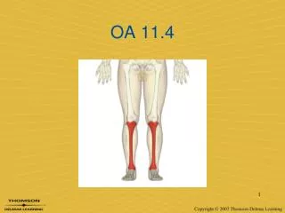
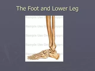
THE ANKLE & LOWER LEG. Lower Leg. Knee cap. Femur. Medial condyle of femur. Lateral condyle of femur. Medial condyle of tibia. Lateral condyle of tibia. Tibial tuberosity. Fibula. Tibia. Lateral malleolus. Medial malleolus. Talus (tarsal bone). Right Leg, Anterior View.

THE ANKLE & LOWER LEG
E N D
Presentation Transcript
Lower Leg Knee cap Femur Medial condyle of femur Lateral condyle of femur Medial condyle of tibia Lateral condyle of tibia Tibial tuberosity Fibula Tibia Lateral malleolus Medial malleolus Talus (tarsal bone) Right Leg, Anterior View Click R Button for Slideshow
Lower Leg2 Knee cap Femur Medial condyle of femur Lateral condyle of femur Medial condyle of tibia Lateral condyle of tibia Tibial tuberosity Fibula Tibia Lateral malleolus Medial malleolus Talus (tarsal bone) Right Leg, Anterior View
Mnemonic for Learning Tarsal Bones: Tiger Cubs Need M I L C Navicular A boat It sails on the Cs Talus Medial cuneiform (1) Intermediate cuneiform (2) Lateral cuneiform (3) Calcaneus Cuboid Click R Button for Slideshow
Foot Quiz H G J F E A C D B Right, Superior View Answers: Next Slide; for Drill Click Back & Forth
Foot Quiz Answers H. Intermediate Cuneiform (2) G. Medial Cuneiform (1) J. Lateral Cuneiform (3) F. Navicular E. Talus A. Phalanges C. Cuboid B. Metatarsals D. Calcaneus Right, Superior View
Talocrural joint • Composed of: • Lateral Malleolus (Distal Portion of Fibula) • Medial Malleolus (Distal portion of Tibia) • Talus
Talocrural joint “cont.” • JOINT MOTIONS: PLANTAR FLEXION DORSIFLEXION
Subtalar Joint • BONES • CALCANEUS • TALUS
SUBTALAR JOINT (Cont.) • JOINT MOTIONS • INVERSION • EVERSION
Distal Tib-Fib Joint • No true motion • Ligaments prevent the tibia and fibula from spreading apart.
LIGAMENTS AND CONNECTIVE TISSUES • INTEROSSEOUS MEMBRANE • CONNECTS TIBIA AND FIBULA
LIGAMENTS AND CONNECTIVE TISSUES • Deltiod ligament • Medial aspect of ankle • Prevents eversion • Very strong rarely torn!
LIGAMENTS • THREE LATERAL LIGAMENTS: • ANTERIOR TALOFIBULAR • POSTERIOR TALOFIBULAR • CALCANEOFIBULAR • PREVENT INVERSION
LIGAMENTS AND CONNECTIVE TISSUES • Anterior and Posterior TIBIOFIBULAR ligaments • BRIDGE THE TIBIA AND FIBULA
MUSCLES OF ANKLE AND LOWER LEG • MUSCLES ON THE ANTERIOR SIDE • DORSIFLEX • EXTENSOR DIGITORUM LONGUS MUSCLE • EXTENSOR HALLUCIS LONGUS MUSCLE • TIBIALIS ANTERIOR MUSCLE
MUSCLES OF ANKLE AND LOWER LEG • SUPERFICIAL POSTERIOR COMPARTMENT • PLANTARFLEX • SOLEUS • GASTROCNEMIUS
MUSCLES OF ANKLE AND LOWER LEG • LATERAL COMPARTMENT • EVERSION • PERONEUS LONGUS • PERONEUS BREVIS
MUSCLES OF ANKLE AND LOWER LEG • DEEP POSTERIOR COMPARTMENT • INVERSION • TIBIALIS POSTERIOR MUSCLE • FLEXOR DIGITORUM LONGUS • FLEXOR HALLUCIS LONGUS
Gastrocnemius • Origin • Medial head medial femoral condyle; • Lateral head lateral condyle • Insertion the Achilles tendon, inserting on the middle 1/3 of the posterior calcaneal surface • Action Powerful plantar flexor of ankle • InnervationTibial nerve (S1, S2)
Soleus • Origin fibular head, Insertion Achilles tendon, inserting on the middle 1/3 of the posterior calcaneal surface • Action Powerful plantar flexor of ankle • Innervation Tibial nerve (S1, S2)
Plantaris • Origin supracondylar line of distal femur • Insertion Middle 1/3 of the posterior calcaneal surface, just medial to Achilles tendon • Action Plantar flexor of ankle; also flexes knee • Innervation Tibial nerve (L5, S1, S2)
Tibialis Posterior • Origin medial fibula, • Insertion 2 • superficial slip inserts on the tuberosity of the navicular bone; • deeper slip plantar sufraces of metatarsals 2 - 4 and second cuneiform • Action Principal invertor of foot • Innervation Tibial nerve (L4, L5)
Flexor Digitorum Longus • Origin Posterior surface of tibia • Insertion Splits into four slips these slips then insert on plantar surface of bases of 2nd - 5th distal phalanges • Action Flexes toes 2 - 5; also helps inversion of ankle • Innervation Tibial nerve
Flexor Hallucis Longus • Origin Inferior 2/3 of posterior surface of fibula, • Insertion Plantar surface of base of distal phalanx of great toe • Action Flexes great toe, helps to invert ankle • Innervation Tibial nerve
Tibialis Anterior • Origin Lateral condyle of tibia, • Insertion 1st cuneiform and on base of first metatarsal • Action Dorsiflexor of ankle • Innervation Deep peroneal nerve (L4, L5, S1)
Extensor Hallucis Longus • Origin Anterior fibula and the interosseous membrane • Insertion Base and dorsal center of distal phalanx of great toe • Action Extends great toe and dorsiflexes ankle • Innervation Deep peroneal nerve (L4, L5, S1)
Extensor Digitorum Longus • Origin head of fibula, • Insertion Splits into 4 tendon slips each of which insert on middle and distal phalanges • Action Extend toes 2 - 5 and dorsiflexes ankle • Innervation Deep peroneal nerve (L4, L5, S1)
Peroneus Tertius • Origin fibular shaft surface • Insertion base of the fifth metatarsal • Action evert the foot • Innervation Deep peroneal nerve
Peroneus Brevis • Origin Inferior 2/3 of lateral fibular surface; • Insertion Base of 5th metatarsal base • Action Everts foot Innervation Superficial peroneal nerve
Peroneus Longus • Origin Head of fibula, • Insertion 1st metatarsal base • Action Everts foot • Innervation Superficial peroneal nerve