Doyle et al., Science 1998; 280: 69
370 likes | 524 Vues
Doyle et al., Science 1998; 280: 69. Jiang et al., Nature 2002 417; 523. Depending on the type of ion channel which opens, the postsynaptic cell membrane becomes either depolarized or hyperpolarized.

Doyle et al., Science 1998; 280: 69
E N D
Presentation Transcript
Doyle et al., Science 1998; 280: 69 Jiang et al., Nature 2002 417; 523
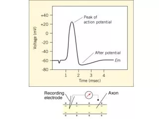
Depending on the type of ion channel which opens, the postsynaptic cell membrane becomes either depolarized or hyperpolarized. • Ions will tend to follow the concentration gradient from high to low concentration, and the electrostatic gradient towards the opposite charge.
INTEGRATION OF EXCITATORY AND INHIBITORY SYNAPSES INPUTS RESPONSE
Inhibitory Post Synaptic Potential Excitatory Post Synaptic Potential IPSP Neuron 2
Postsynaptic neuron Inhibitory neurotrans-mission prevents excitation of the post-synaptic neuron Inhibitory GABA presynaptic neuron excitatory presynaptic neuron inhibitory
Fig. 5 Signal transduction pathways of G-protein coupled receptors. Upon activation of the G-protein coupled receptor (GPCR) by the binding of an appropriate ligand (L), the associated heterotrimeric G-protein, which contains an α, β and γ subunit, exchang... Steven J. Husson , Inge Mertens , Tom Janssen , Marleen Lindemans , Liliane Schoofs Neuropeptidergic signaling in the nematode Caenorhabditis elegans Progress in Neurobiology Volume 82, Issue 1 2007 33 - 55 http://dx.doi.org/10.1016/j.pneurobio.2007.01.006
Fig. 5 Signal transduction pathways of G-protein coupled receptors. Upon activation of the G-protein coupled receptor (GPCR) by the binding of an appropriate ligand (L), the associated heterotrimeric G-protein, which contains an α, β and γ subunit, exchang... Steven J. Husson , Inge Mertens , Tom Janssen , Marleen Lindemans , Liliane Schoofs Neuropeptidergic signaling in the nematode Caenorhabditiselegans Progress in Neurobiology Volume 82, Issue 1 2007 33 - 55 http://dx.doi.org/10.1016/j.pneurobio.2007.01.006
Glutamate fast neurotransmission Synthesis, packaging, reuptake, degradation (error - should be EAAT)
Pharmacology AMPA agonists: AMPA, glutamate antagonists: CNQX, NBQX Kainate agonists: kainic acid, glutamate antagonist: CNQX NMDA agonists: glutamate, aspartate, NMDA antagonists: D-APV, D-AP5, MK-801, Ketamine, Phencyclidine, (Mg++)
Pharmacology AMPA agonists: AMPA, glutamate antagonists: CNQX, NBQX Kainate agonists: kainic acid, glutamate antagonist: CNQX NMDA agonists: glutamate, aspartate, NMDA antagonists: D-APV, D-AP5, MK-801, Ketamine, Phencyclidine, (Mg++)
NMDA receptors show slow onset and decay kinetics Some synapses have both glutamate receptor types, and produce a two-component synaptic current
If the time interval between Ca2+ spikes is long compared to the dissociation time of Ca2+–calmodulin (low frequency), initial binding is presumed to lead to a separation of kinase domains; however, transphosphorylation of Thr286 does not occur before Ca2+–calmodulin dissociates. If the interval between Ca2+ spikes is short compared to the Ca2+–calmodulin dissociation time (high-frequency regime) Ca2+–calmodulin remains bound to the first pair of kinase domains long enough for a second, slow step to occur, in which the activated kinase domains capture the regulatory segments of adjacent kinase domains. This potentiates the binding of Ca2+–calmodulin to those domains with increased affinity, resulting in the phosphorylation of Thr286 and acquisition of autonomy (Ca2+–calmodulin-independent activity). Caho …. Kuriyan (2010) Nature Structural & Molecular Biology17,264–272(2010)
Cooling activates TRPM8 by shifting the voltage dependence of activation. a, Whole-cell TRPM8 currents at +100 and -80 mV in response to slow cooling of the bath solution. b, Normalized current responses at +100 and -80 mV in function of temperature. Voets et al (2004) Nature 430: 748 - 54
Heating activates TRPV1 by shifting the voltage dependence of activation. c, Current traces at different temperatures in response to voltage steps ranging from 2120 to þ160 mV. d, Steady-state activation curves at different temperatures for the currents shown in c. Lines represent Boltzmann functions fitted to the data. e, V 1/2 as a function of temperature (n values between 4 and 9). Dashed line represents the change in V 1/2 as predicted by the two-state model (see text). Voets et al (2004) Nature 430: 748 - 54