Download
1 / 35
380 likes | 790 Vues
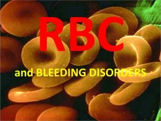
RBC Morphology and Cases. Dr. Gilbert’s Lab. CASE 1. Normal peripheral blood smear A normal complete blood count (CBC) and normal peripheral blood smear are shown. Slide 1.1 A CBC demonstrates normal findings.

E N D
RBC Morphology and Cases Dr. Gilbert’s Lab
CASE 1 Normal peripheral blood smear • A normal complete blood count (CBC) and normal peripheral blood smear are shown.
Slide 1.2A normal peripheral blood smear is seen here with a normal segmented neutrophil and a lymphocyte.
Questions • Define the terms and explain the values and the reference ranges. • Demonstrate normal WBC, RBC, and platelet morphology. • Demonstrate how to do a WBC differential count. • Demonstrate how to estimate platelet counts. With a 100X oil immersion objective, 5 to 20 platelets should be found in each field examined under the microscope on a smear from a patient with a normal platelet count.
CASE 2 • History: • A 72 year old male has the findings shown below on a CBC. The peripheral blood smear shows red blood cells with hypochromasia and microcytosis.
Slide 2.2Note the poikilocytosis and microcytosis and hypochromia in this peripheral blood smear.
Questions • Demonstrate how to estimate the red blood cell MCV. A normal red blood cell is 2/3 the size of a small lymphocyte, or about the size of (or slightly smaller than) the lymphocyte nucleus. • Demonstrate the appearance of hypochromic RBC's. • What is the diagnosis from these findings? • Which of the following tests would be most useful to determine the etiology: A. Hemoglobin electrophoresis B. Reticulocyte count C. Stool for occult blood D. Vitamin B12 assay E. Bone marrow biopsy
CASE 2: Iron deficiency anemia • What is the diagnosis from these findings? Hypochromic microcytic anemia (from probable iron deficiency). • Which of the following tests would be most useful to determine the etiology: A. Hemoglobin electrophoresis B. Reticulocyte count C. Stool for occult blood D. Vitamin B12 assay E. Bone marrow biopsy Answer: C This patient most likely has a blood loss anemia, and a colon cancer is a likely source in an older male. Laboratory testing consistent with iron deficiency anemia would include a low serum ferritin and low serum iron with low % saturation.
CASE 3 • History: • A 48 year old male has become progressively more fatigued at the end of the day. This has been going on for months. In the past month he has noted paresthesias with numbness in his hands. A CBC demonstrates the findings shown below. A peripheral blood smear (the slide is representative of this condition) shows red blood cells displaying macro-ovalocytosis and neutrophils with hypersegmentation.
Slide 3.2A peripheral blood smear demonstrates megaloblastic changes. Describe the appearance of the neutrophil.
Questions • Describe macrocytosis and macro-ovalocytosis. • Describe hypersegmented PMN's. • What is the diagnosis from these findings? • Which of the following tests would be most useful to determine the etiology: A. Hemoglobin electrophoresis B. Reticulocyte count C. Stool for occult blood D. Vitamin B12 assay E. Bone marrow biopsy 3. How do you explain the neurologic findings?
CASE 3: Megaloblastic anemia • What is the diagnosis from these findings? This is a macrocytic (megaloblastic) anemia. The neurologic findings suggest vitamin B12 deficiency (pernicious anemia). • Which of the following tests would be most useful to determine the etiology: A. Hemoglobin electrophoresis B. Reticulocyte count C. Stool for occult blood D. Vitamin B12 assay E. Bone marrow biopsy Answer: D Macrocytic anemia could also be caused by a folate deficiency, but the neurologic findings would not be present. 3. How do you explain the neurologic findings? The B12 deficiency leads to a subacute combined degeneration of the spinal cord (posterior and lateral columns).
CASE 4 • History: • A 30 year old female has sudden onset of fever, abdominal pain, tachycardia, nausea, and jaundice. She is anemic, with Hgb 11.1 g/dL, Hct 28.8%, and MCV 77 fL. The WBC count is normal, and she has marked reticulocytosis. RBC morphology shows small cells that lack central pallor. Physical examination reveals a palpable spleen tip. A month ago, a CBC showed the following findings:
Slide 4.1A CBC is shown here characteristic for this condition.
Slide 4.2A peripheral blood smear is shown here. Compare the size of the RBC's to the lymphocyte nucleus.
Questions • Describe spherocytes and discuss the differential diagnosis. • Discuss the terms reticulocytosis and polychromasia. • What is the diagnosis from these findings? • The probable association(s) with this disease is(are): A. Autosomal dominant, European ancestry B. X-linked, Asian ancestry C. Autosomal recessive, Middle Eastern ancestry D. Autosomal recessive, West African ancestry E. Sporadic occurrence 3. An aplastic crisis in this patient could be initiated by: A. Quinacrine B. Parvovirus infection C. Decreased oxygen tension D. Exposure to cold E. Transfusion
CASE 4: Hereditary spherocytosis • What is the diagnosis from these findings? Hereditary spherocytosis. • The probable association(s) with this disease is(are): A. Autosomal dominant, European ancestry B. X-linked, Asian ancestry C. Autosomal recessive, Middle Eastern ancestry D. Autosomal recessive, West African ancestry E. Sporadic occurrence Answer: A 3. An aplastic crisis in this patient could be initiated by: A. Quinacrine B. Parvovirus infection C. Decreased oxygen tension D. Exposure to cold E. Transfusion Answer: B Parvovirus (sometimes called "fifth disease") infects RBC precursors.
CASE 5 FYI • History: • A 29 year old female with a diagnosis of acute promyelocytic leukemia develops petechiae and ecchymoses at multiple sites. She becomes progressively more hypotensive before losing consciousness. A CBC shows Hgb 8.7 g/dL, Hct 24.5%, MCV 85 fL, platelets 17,000/microliter, and WBC count 8700/microliter. The peripheral blood smear demonstrates many fragmented RBC's. • Demonstrate schistocytes and helmet cells
Slide 5.1Note the fragmented RBC's in this peripheral blood smear.
Questions • What is the diagnosis from these findings? • Discuss the conditions in which this can occur. • Which of the following tests would be most useful to diagnose this condition in this patient with acute promyelocytic leukemia: A. Hemoglobin electrophoresis B. Reticulocyte count C. Coagulation tests D. Vitamin B12 assay E. Bone marrow biopsy
Additional History: • For comparison, a CBC from a patient with a malfunctioning mechanical heart valve is shown below. Spot the key feature in the RBC histogram.
Slide 5.2Note the high red cell distribution width in this CBC.
CASE 5: Microangiopathic hemolytic anemia • What is the diagnosis from these findings? Microangiopathic hemolytic anemia, in this case disseminated intravascular coagulation (DIC). Note: this slide does not show the leukemia, only DIC. • Discuss the conditions in which this can occur. Gram-negative sepsis with endotoxemia, disasters that release tissue thromboblastic substances (obstetric complications such as abruptio placenta, major trauma, malignancies), and promyelocytic leukemia. • Which of the following tests would be most useful to diagnose this condition in this patient with acute promyelocytic leukemia: A. Hemoglobin electrophoresis B. Reticulocyte count C. Coagulation tests D. Vitamin B12 assay E. Bone marrow biopsy Answer: C The D-dimer test would be the most specific. Additional History: • For comparison, a CBC from a patient with a malfunctioning mechanical heart valve is shown. Spot the key feature in the RBC histogram. There is hemolysis, so the RDW (red cell distribution width) is markedly increased.
CASE 6 • History: • A 20 year old African-American male comes to the emergency room because of the onset of severe abdominal pain. A CBC shows Hgb 4.8 g/dL, Hct 12.8%, MCV 80 fL, platelets 205,000/microliter, and WBC count 9800/microliter with differential count showing 12 NRBC's per 100 WBC's. Examination of the peripheral smear shows marked poikilocytosis and anisocytosis with numerous sickled erythrocytes.
Slide 6.1Note the shape of the RBC's in this peripheral blood smear.
Slide 6.2Note the shape of the RBC's in this peripheral blood smear.
Questions • Demonstrate the sickled erythrocytes • Discuss the demographics of this disease • What is the diagnosis from these findings? • Which of the following tests would be most useful to determine the etiology: A. Hemoglobin electrophoresis B. Reticulocyte count C. Stool for occult blood D. Vitamin B12 assay E. Bone marrow biopsy 3. The "crisis" in this patient was initiated by: A. Quinacrine ingestion B. Parvovirus infection C. Decreased oxygen tension D. Exposure to cold E. Transfusion therapy
CASE 6: Sickle cell anemia • What is the diagnosis from these findings? Sickle cell anemia (Hgb SS). • Which of the following tests would be most useful to determine the etiology: A. Hemoglobin electrophoresis B. Reticulocyte count C. Stool for occult blood D. Vitamin B12 assay E. Bone marrow biopsy Answer: A 3. The "crisis" in this patient was initiated by: A. Quinacrine ingestion B. Parvovirus infection C. Decreased oxygen tension D. Exposure to cold E. Transfusion therapy Answer: C This is why people with this disease do not live in Utah.
CASE 7 • History: • This CBC demonstrates the findings following treatment for one of the conditions given above.
Slide 7.1Note the double peak to the histogram indicating RBC size.
Question • Can you determine what that treatment was?
CASE 7: Transfusion • This CBC demonstrates the findings following treatment for one of the conditions given above. • Can you determine what that treatment was? The RBC histogram demonstrates a dual population of RBC's following transfusion.