Design and Background Correction in Spectrophotometry: Understanding Detector Mechanisms
This lecture delves into the principles and designs of spectrophotometers, focusing on detectors, background correction methods, and spectral peak characteristics. Key topics include the Harris Monochromator, various light sources, phototubes, and photomultiplier setups, including their efficiency and response to light at different wavelengths. It discusses the significance of signal processing techniques, the impact of noise on spectral data, and methodologies for eliminating background interference. A comprehensive overview of quantum states, molecular versus atomic spectra, and the Doppler effect in wavelength measurements is also presented.

Design and Background Correction in Spectrophotometry: Understanding Detector Mechanisms
E N D
Presentation Transcript
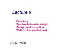
Lecture 8 Detectors Spectrophotometer design Background correction Width of the spectral peak Ch. 20 Harris
Monochromator (filter, wavelength selector) Light Source Detector Sample Spectrometer Data Processing
A phototube K/Cs/Sb GaAs - + Ag/O/Cs 90 V Wavelength, nm
Photomultiplier Several electrons for each incident electron Several electrons for each photon Quarz envelope Light, h Photoemissive cathode Anode
Photoelectromultiplier dynode dynode electron photon
Single beam Double beam
Single beam design with 210-element area detector
Flat background Ipeak I Ipeak – I0 Intensities at two different wavelengthes Peak I0 Baseline wavelength
Flat background Ipeak I constant Peak First derivative eliminates flat background I0 Baseline Wavelength (l)
Linear Background Ipeak I Ipeak – (I1+I2) / 2 I1 Background I2 Wavelength (l)
Linear Background I Ibackground = Kl+b First: Background Second: Second derivative eliminates linear background Wavelength (l)
Original Second 4th First
Well defined peaks and valleys High noise Some peaks visible Almost featureless: little information Low noise
Only photons of one specified wavelength are absorbed Excited state Ground state
Width Velocity of a particle >1000 m/s Doppler Speed of light 300,000,000 m/s collisions Approximately as Doppler Both effects increase with the temperature and decrease with the size of the molecule/atom Width is around 0.01-0.001 nm in visible area Multiple energy levels
Energy levels of excited state Energy sublevels Energy levels of ground state
A primitive rule: Molecular spectra - widebands Atomic spectra - narrowlines