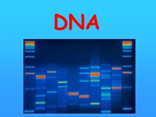
DNA
Explore pivotal experiments and discoveries that established DNA as the hereditary material, from Griffith's transformation experiment to Chargaff's rules and Watson and Crick's DNA structure elucidation. Discover how Franklin's X-ray crystallography images were instrumental in unraveling the DNA double helix.

DNA
E N D
Presentation Transcript
DNA Molecular Genetics 7.1
Understandings: • Nucleosomes help to supercoil the DNA. • DNA structure suggested a mechanism for DNA replication. • DNA polymerases can only add nucleotides to the 3’ end of a primer. • DNA replication is continuous on the leading strand and discontinuous on the lagging strand. • DNA replication is carried out by a complex system of enzymes. • Some regions of DNA do not code for proteins but have other important functions. Applications and skills: • Application: Rosalind Franklin’s and Maurice Wilkins’ investigation of DNA structure by X-ray diffraction. • Application: Use of nucleotides containing dideoxyribonucleic acid to stop DNA replication in preparation of samples for base sequencing. • Application: Tandem repeats are used in DNA profiling. • Skill: Analysis of results of the Hershey and Chase experiment providing evidence that DNA is the genetic material. • Skill: Utilization of molecular visualization software to analyze the association between protein and DNA within a nucleosome.
I. The Chemical Nature of Nucleic Acids. How did we come to believe that DNA was the hereditary material? DNA was established as the hereditary material over the other candidate molecule, protein, by a series of experiments. a) Frederick Griffith (1928). Used mice and strains of the bacteria,Streptococcus pneumoniae. Was able to transform non-pathogenic strain of bacteria into pathogenic form by growing them with heat-killed pathogenic bacterial cells. Transformation was heritable. Non-pathogenic trait had acquired something from heat-killed bacteria.
History of DNA as hereditary material • Avery, MacLeod, and McCarty (1944) showed that the transforming agent was DNA.
Hershey and Chase • Hershey and Chase (1952) using viruses that infect bacteria (bacteriophage) of E.coli, a gut bacteria, showed that it was the DNA component that was transferred during infection and was responsible for reprogramming the host bacterial cell. • Radioactively labeled sulfur (S) which is found in proteins to see if proteins were transferred from the bacteriophage to the bacteria • Radioactively labeled phosphorus (P) which is found in DNA to see if DNA was transferred
Hershey and Chase Experiment: Evidence that DNA is hereditary material
Hershey and Chase Experiment • What they found was the radioactive phosphorus was transferred from the bacteriophage to the bacteria • This provided information that DNA was the genetic material
DNA as hereditary material • Erwin Chargaff in 1947 analyzed DNA composition of different organisms, including eukaryotes. He found that DNA composition was species specific. The amount and ratios of nitrogenous bases vary from one species to another. In every species he looked at there was a regularity in base ratios: Number of adenine (A) residues approximately equaled the number of thymine (T), and the number of guanines (G) equaled the number of cytosine ( C). Chargaff’s rules: A=T and G=C (1950)
More DNA History e) Watson and Crick published in 1953 in the British journal Nature, the structure of DNA. It obeyed Chargaff’s rules and explained the X-Ray photos produced by Rosalind Franklin and Maurice Wilkins.
Rosalind Franklin (1920-1958) • Franklin was an English chemistry and worked on X-ray crystallography • While at King’s College in London she worked on X-ray diffraction images of DNA • This lead to the discovery of the DNA double helix • She also found that the double helix depends on the level of hydration and discovered A-DNA and B-DNA • Her colleague Maurice Wilkins shared this information with Watson and Crick • While Watson and Crick are credited with discovering the double helical structure of DNA, Franklin was the first to find the structure • She died of ovarian cancer at 37 (most likely due to radiation) • She most likely would have won the Nobel Prize along with Watson and Crick but the award cannot be awarded posthumously
Watson and Crick and their use of model making on the elucidation of the structure of DNA • In the 1950s the race to find the structure of DNA was on. Many scientists were working to discover what the shape of DNA might look like. With the discovery by Chargaffof the base-pairing nature of nucleic acids and Pauling’s work on protein structure, Francis Crick and James Watson were intrigued in deducing the structure of DNA. While Franklin and Wilkins took an experimental approach, Watson and Crick took a physical approach at Cambridge University using models. After attending a lecture by Franklin they tried to use her data to construct a model using cardboard cut outs in the labs. This model failed and their research came to a stop. When Franklin discovered the x-ray crystallography of the DNA molecule Watson and Crick were able to use this data to begin construction of the DNA molecule again in 1953.
II. Watson and Crick Model • DNA is a double helix with the bases of two strands pointed inward toward each other, forming base-pairs. • Base-pairs always consist of purines which are large, pointing toward pyrimidines, which are small, keeping the diameter of the molecule at 2 nanometers. • Double helix is stabilized as a duplex DNA molecule composed of two antiparallel strands, one chain running 3’ to 5’ and the other 5’ to 3’. • Watson and Crick were awarded the Nobel prize in physiology/medicine in 1962 (after Franklin’s death)
Nucleotide Simplified IB wants you to represent a nucleotide similar to this!
Watson and Crick model cont. • An A-T pair has 2 hydrogen bonds while G-C base-pair has 3. • Sugar-phosphate chains are on the outside which allows the more hydrophobic nitrogenous bases to swivel to the interior away from the aqueous medium. • Model suggests the general mechanisms for DNA replication. Strands that are not identical but are complimentary, serve as templates for the other stands.
Important features of nucleic acids and DNA • DNA contains 3 main components: 1) the sugar deoxyribose which is a pentose. 2. Phosphate group 3. Nitrogen containing bases called Purines (A, G), Pyrimidines (T, C). RNA contains uracil instead of thymine. Each nucleotide consists of sugar attached to a phosphate group and a base. • Carbons of the sugar are numbered 1’ to 5’ proceeding clockwise from the O atom. They are ‘(prime) to distinguish the carbons in a sugar from those in a base.
DNA/RNA features • Phosphate group is attached to the 5’ carbon atom of the sugar and the base is attached to the 1’ carbon atom. A free hydroxyl is attached to the 3’ carbon atom. • The 5’ phosphate and 3’ hydroxyl groups allow DNA and RNA to form long chains of nucleotides, because these 2 groups can react chemically with each other.
DNA synthesis continued • The result is a dehydration synthesis, eliminating water and forming a covalent bond that links the 2 groups. The linkage is called a phosphodiester bond. The 5’ and 3’ ends still available to link to other nucleotides. • Synthesis requires energy. This is provided by the nucleoside triphosphates. The cleavage of the triphosphate to a monophosphate produces the energy necessary to form the phosphodiester bond. • DNA has directionality. Sequence of bases usually expressed in the 5’ to 3’ direction.
III. DNA Replication Watson and Crick model for DNA structure suggested a template mechanism for DNA replication. Steps proposed: • Two DNA strands separate • Each strand is a template for assembling a complimentary strand. • Nucleotides line up singly along the template strand in accordance with the base-pairing rules (A-T and G-C). • Enzymes link the nucleotides together at their sugar-phosphate groups.
Proposed DNA Replication Mechanism: Semi-conservative • Proposed that DNA replication is semi-conservative. This means that each of the two daughter molecules produced through replication will have one old or conserved strand from the parent molecule and one newly created strand. • Meselson and Stahl (1958) of Cal Tech grew bacteria in a medium containing the heavy isotope of nitrogen, 15N, which became incorporated into bases of the bacterial DNA. Provided evidence demonstrating semi-conservative replication.
A. Process of DNA replication • Is complex. The helical molecule must untwist while it copies its two antiparallel strands simultaneously. Requires the cooperation of over a dozen enzymes and other proteins. • Is extremely rapid. In prokaryotes, up to 500 nucleotides are added per second. It takes only a few hours to copy the 6 billion bases of a single human cell. • Is accurate. Only about 1 in a billion nucleotides are incorrectly paired.
DNA Replication Proteins of E. coliBegan 1960: Arthur Kornberg • Helicase: Unwinds the double helix • Topoisomerase I: Relieves torque by making small cuts in DNA. (Called DNA Gyrase in prokaryotes) • RNA primase: Synthesizes RNA primers • RNase H: removes the RNA primer made by primase. Not considered in IB analysis of Prokaryotic DNA Rep. because necessary only in Eukaryotes). • DNAPolymerase I(discovered by Arthur Kornberg while he was Washington Univ.) Fills in gaps of DNA and excises the primer in Prokaryotes. • DNAPolymerase III: Major DNA polymerase that synthesizes DNA at the 3’ end. IT CAN ONLY ADD TO THE 3’ END! • DNA Ligase: Joins the ends of DNA segments • Single-stranded binding protein: stabilizes single-stranded regions • Initiator proteins needed to find the ORI or origin of replication
B. DNA Polymerase III • True replicating enzyme. • Contains 10 different kinds of polypeptide chains. • It is a dimer, with 2 similar multisubunit complexes. Each complex catalyzes the replication of 1 strand of DNA • a subunit: catalyzes addition of nucleotides to a growing chain. • b subunit: proofreads for mistakes • Ring shaped B dimer that clamps the polymerase III complex around the DNA double helix. Polymerase III progressively threads the DNA through the enzyme complex, moving it at a rapid rate.
The Need for a Primer • DNA Polymerase III can add nucleotides only to a chain of nucleotides that is already paired with the parent strands. It therefore cannot link the first nucleotides in a newly synthesized strand. • This requires an RNA polymerase called Primase to construct an RNA primer. DNA Polymerase III recognizes the primer and adds DNA nucleotides to it to construct the DNA strands. • The RNA nucleotides in the primers are then replaced by DNA molecules. • The promoter region of DNA in an example of non-coding DNA with a function
D. The two strands are assembled in different ways • DNA Polymerase III can add nucleotides only to the 3’ end of a DNA strand (the end with the –OH group attached to a 3’carbon atom). Replication always proceeds in the 5’ to 3’ direction on a growing DNA strand. Since 2 parent strands of a DNA molecule are antiparallel, the new strands are oriented in opposite directions along the parent templates at each replication form. • New strands must be elongated by different mechanisms.
1. Leading Strand • Elongates towards the replication fork; is built up simply by adding nucleotides continuously to its growing 3’ end. 5’ to 3’
2. Lagging Strand • Elongates away from the replication fork • Is synthesized discontinuously as a series of short segments that are later connected. • These segments are called Okazaki fragments, after Reiji Okazaki at Nagoya University who discovered them and proposed the leading/lagging strand models. • Okazaki fragments are about 100-200 nucleotides long in eukaryotes because of multiple initiation sites and 1000-2000 nucleotides in prokaryotes.
Lagging Strand synthesis • Okazaki fragments are synthesized by DNA polymerase III in the 5’ to 3’ direction, beginning at replication fork and moving away from it. • When polymerase reaches the 5’ end of the lagging strand, another enzyme, DNA ligase, attaches the fragment to the lagging strand. • DNA is further unwound, new RNAprimers are constructed, and DNA polymerase III jumps ahead toward the replication fork to construct another Okazaki fragment.
E. The Replication Process takes place in 5 interlocking steps Step 1. Opening up theDNA double helix. DNA must be opened up and separated. • Binding of initiator proteins to the origin of replication (ori) • Unwinding the duplex involves enzyme called helicase binding to and moving along one strand, shouldering aside the other strand as they go. • Unwound portion of DNA double helix is stabilized by single-stranded binding proteins which bind and protect them from cleavage and rewinding. • Relieving the torque generated by unwinding: topoisomerase cleaves a strand of the helix, allowing it to swivel around the intact strand, and then reseal the broken strand.