Initial ventilator settings
Dr Chaitanya Vemuri Int.Med M.D Trainee. Initial ventilator settings. The choice of ventilator settings – guided by clearly defined therapeutic end points. In most of cases : primary goal is to correct abnormalities of arterial blood gas tensions Accomplished by

Initial ventilator settings
E N D
Presentation Transcript
Dr ChaitanyaVemuri Int.Med M.D Trainee Initial ventilator settings
The choice of ventilator settings – guided by clearly defined therapeutic end points. • In most of cases : primary goal is to correct abnormalities of arterial blood gas tensions • Accomplished by • adjusting minute volume - to correct hypercapnea • oxygen supplementation – to correct hypoxemia Introduction
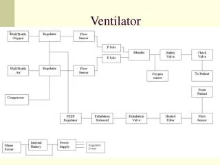
Choice of inspired gas composition Means to ensure the machine’s sensing of patient’s demand Definition of machine’s mechanical output General aspects of ventilator management
Confirm indication for mechanical ventilation • Invasive / Non invasive • Check Connections & Circuit • Self test • Select mode • Set variables • Alarm settings • Connect to patient • Monitor and reassess
Patient not breathing Patient breathing but not enough Patient breathing enough, but pt hypoxemic / hypercapneic Patient breathing with normal gas exchange, but working hard Airway protection indications
LABORATORY CRITERIA CLINICAL CRITERIA OTHER CRITERIA Indications
Blood gases : PaO2 < 55 mm Hg PaCo2 > 50 mm Hg pH < 7.32 PFT : Vital Capacity < 10 ml/Kg -ve inspiratory force <25cm H20 FEV 1 < 10 mL/Kg LABORATORY CRITERIA
Apnea / Hypopnea Respiratory distress with altered mentation Clinically apparent increasing work of breathing unrelieved by other interventions Need for airway protection Clinical criteria
Controlled hyperventilation ( eg head injury ) • Severe circulatory shock • THERE IS NO ABSOLUTE CONTRAINDICATION FOR MECHANICAL VENTILATION Other criteria
To check : - leak - compliance - resistance of circuit - sensors • Needs to be done : - before connecting to patient - once in 2 weeks - whenever circuit is changed Self test
Depends on : Patients requirement User comfort Availability Select mode
For PO2 : adjust FiO2, PEEP For PCo2 : adjust TV , RR Basic principles
Tailored to need of the patient SIMV / A/C – versatile modes for initial settings In pts with good resp drive & mild – mod resp failure – PSV MODE OF VENTILATION
Initial TV : 5 – 8 ml/Kg of ideal bd wt Lowest values are recommended in presence of Obstructive airway ds & ARDS Goal : to adjust TV so that plateau pressures are less than 35 cm H20 Tidal volume
8 – 12 breaths per minute : pts not requiring hyperventilation for treatment of toxic/metabolic acidosis or intracranial injury Initial rate may be low ( 5 – 6 breaths per min ) in asthmatic pts where permissive hypercapnic technique is used Respiratory rate
Lowest FiO2 that produces an Sp02 > 90 % PaO2 > 60 mm Hg is recommended Supplemental o2 therapy
Normal I:E ratio to start is 1:2 Reduced to 1:4 or 1:5 in presence of obstructive airway disease in order to air trapping Inverse I:E – in ARDS Inspiration : expiration ratio
60 L/min is typically used Increased to 100 L/min : to deliver TVs quickly and allow for prolonged expiration in presence of obstructive airway ds INSPIRATORY FLOW RATE
Titrated according to PEEP and BP • High PEEP ( > 10 H20 ) – pneumonia, ards • PEEP – reduces risk of atelectasis - increase no of open alveoli ( decrease V/Q mismatch ) - in CHF : decrease venous return • Physiological PEEP ( 3-5 cm H20 ) : to prevent decrease in FRC in normal lungs Positive end expiratory pressure ( peep )
Set at -1 to -2 cm H20 NEWER VENTILATORS SENSE INSPIRATORY FLOW and thereby reduce work of breathing associated with ventilator triggering Sensitivity ( TRIGGER )
Mode : Complete / Partial . VCV/PCV • Rate : titrate to Pco2 • Tidal Volume : 5 – 8 ml / Kg • Flow rate & Pattern : 4 – 8 times Minute Ventilation • I:E = 1:2 to 1:4 • FiO2 : titrate to O2 Saturation / Pa O2 • PEEP : titrate to PaO2 & BP • Trigger : Adjust to synchronize SET VARIABLES
Fixed alarms : disconnection o2 sensor Set alarms : volume pressure rate apnea Alarm settings
Patient • Monitor : pulse , bp , rr, spO2 • Ventilator • Abg • Volume • Pressure • Rate • Patient comfort / synchrony Monitor & reassess
Ventilatory settings in various diseases
For Paralysed pts : CMV or A/C mode For Non paralysed pts : SIMV mode Pts with normal resp effort mild resp failure : PSV mode
Hypoxia corrected by High FiO2 • Increase Expiratory Flow Time to max : to prevent increase intrinsicPEEP • RR : 6 -8 breaths / min ( permissive hypercapnia ) • I : E : increased 1:2 Asthma & copd
A/C mode Tidal Volume : 6 ml/Kg PEEP : 5 Ventilatory rate : 12 titrated to maintain Ph > 7.25 ards
Respond well to positive pressure ventilation (opens alveoli, reduces preload) • Many benefit from trial of noninvasive CPAP / BiPAP • Intubated pts usually manage to oxygenate well • But PEEP can be increased to improve oxygenation and reduce preload Chf