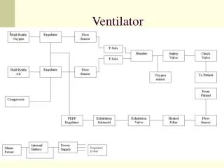
Ventilator Basics
This guide aims to provide a comprehensive understanding of volume-preset mode ventilation in critical care. Learn the differences between Assist Control (AC) and Synchronized Intermittent Mandatory Ventilation (SIMV), the significance of plateau and peak pressures, and how to use these parameters as vital signs in ventilated patients. Gain insights into diagnosing airway obstruction through expiratory waveforms, measuring auto-PEEP, and optimizing ventilatory settings to enhance patient outcomes, especially in cases like ARDS.

Ventilator Basics
E N D
Presentation Transcript
Goals • Understand volume-preset mode of ventilation • Understand the difference between SIMV and AC • Understand the meaning of plateau pressure and peak pressure in volume mode ventilation • Learn how to use peak pressure and plateau pressure as additional “vital signs” in a ventilated patient • Learn how use interpret expiratory waveform to diagnose obstruction in a patient on a ventilator • Learn how to measure auto-PEEP and to decrease auto-PEEP
Volume-Preset Mode Ventilation • Volume-preset mode ventilation are modes where the tidal volume is determined by the clinician • In comparison, pressure-preset mode ventilation are modes where the pressure is specified by the clinician. • In pressure-preset model ventilation, the tidal volume that the patient receives is determined by mechanics of the patient’s lung and airways in response to the pressure specified by the clinician • E.g., In non-compliant lungs, a given pressure setting would result in less tidal volume delivered to the patient
Volume-Preset Mode Ventilation • We will focus exclusively on volume-preset mode ventilation • The primary trials in ARDS were done in volume-present mode ventilation • It is easier to measure mechanical properties of the respiratory system • Measurement of peak pressure and plateau pressure • It is easier to understand how to manipulate the ventilator
Volume-Preset Mode Ventilation • AC = Assist Control • Tidal volume is set by the clinician as well as the respiratory rate • Control means that the ventilator delivers the tidal volume at the set respiratory rate • Any additional breaths over the respiratory rate set by the clinician is guaranteed to be the same tidal volume set by the clinician
Volume-Preset Mode Ventilation • AC = Assist Control • Example: AC with tidal volume 450 mL, respiratory rate 24 breaths/minute • In a paralyzed patient, the patient receives 24 breaths/minute with each breath at a tidal volume of 450 mL • In a non-paralyzed patient with the same ventilator setting, the patient is forced to breathe 24 breaths/minute at a tidal volume of 450 mL. • If a patient breathes above the rate of 24 breaths/minute, each of those additional breaths are also guaranteed to be at 450 mL
Volume-Preset Mode Ventilation • SIMV = Synchronized intermittent mandatory ventilation • Tidal volume is set by the clinician as well as the respiratory rate • Any additional breaths over the respiratory rate set by the clinician is not guaranteed to be the same tidal volume set by the clinician • The tidal volume of the additional breaths are dependent on patient effort • Because the extra breaths are dependent on patient effort, this mode of ventilation is not recommended in patients with sepsis/ARDS because they put additional strain on the patient
Volume-Preset Mode Ventilation • SIMV = Synchronized intermittent mandatory ventilation • Example: SIMV with tidal volume 450 mL, respiratory rate 24 breaths/minute • In a paralyzed patient, the patient receives 24 breaths per minute with each breath at a tidal volume of 450 mL • In a non-paralyzed patient with the same ventilator setting, the patient is guaranteed 24 breaths/minute with a tidal volume of 450 mL during those mandatory breaths • However, if a patient breathes above the rate of 24 breaths/minute, each of those additional breaths are not guaranteed to be at 450 mL and the tidal volume generated depends on the patient’s effort
Oxygenation • To improve the oxygenation of the patient on a ventilator, you can either increase the FiO2 or the PEEP • Increase FiO2 • Increased FiO2 helps to increase oxygenation by increasing the oxygen gradient between the air in the alveoli and the blood • Increase PEEP • PEEP helps to recruit alveoli, thus helping to improve oxygenation • This is important in ARDS • A caveat is that too high of PEEP can potentially lower venous return and as a result, stroke volume and cardiac output thus causing hypotension
Ventilation • To adjust the ventilation of the patient on a ventilator, you can either increase the tidal volume or the respiratory rate • Increase tidal volume • A caveat is that this may increase the plateau pressure • In general, in ARDS, it is essential to decrease the tidal volume if the plateau pressure is > 30 cm H2O • Refer to ARDS lecture slides and subsequent discussion here • Increase respiratory rate • A caveat is that if the patient is already breathing above the respiratory rate that you are setting on the ventilator, then ventilation will not be improved
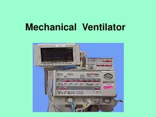
Volume Preset Modes • Examples: • AC (assist control) • SIMV (synchronized intermittent mandatory ventilation) • Important Points: • Tidal Volume set • Flow rate set • Pressure in the system develops in response to the volume pushed in by the ventilator
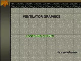
Volume Preset Modes • Refer to the pressure-time graph (top graph) for this discussion • Pressure response depends on the respiratory system • In patients with a very stiff lung, the peak pressure for a given tidal volume will be higher • Similarly, in patients with very tight airways (high resistance), the peak pressure for a given tidal volume will be higher • Note that pressure increases during the inspiratory phase when the ventilator pushes in the tidal volume set by the clinician • This is the active phase of ventilator where the ventilator pushes in the tidal volume specified by the clinician
Volume Preset Modes • Refer to the flow-time graph (bottom graph) • Note that the expiratory phase is passive • The flow drops to zero quickly during the expiratory phase of ventilation • Remember this as we will see what obstruction looks like in the expiratory phase of ventilation
Pressure Response • Given a specific tidal volume, the pressure it takes to overcome the respiratory system is equal to the pressure needed to overcome the resistance of the airways (Presist) and the pressure it takes to expand the alveoli against the elastic recoil of the lung and the chest wall (Pelast) • Total pressure (Ppeak) = Presist + Pelast+ PEEP • This is the total pressure measured at the end of inspiration • Assuming the patient is not actively breathing against the ventilator • The patient should be passive during this measurement otherwise it will not be accurate • For the following discussion, we will leave the PEEP out for simplicity
Pressure Response • Total pressure (Ppeak) = Presist + Pelast + PEEP • We are leaving PEEP out during this discussion for convenience • Presist gives you insight into the resistance of the airways in the respiratory system • Presist depends on flow, according to Ohm’s law • Presist = flow x resistance • Pelast gives you insight into how difficult it is to inflate the alveoli
Pressure Response • Total pressure (Ppeak) = Presist + Pelast • This is assuming that there is no PEEP Presist Pelast • On a ventilator, we can measure Ppeak and Pplat • Pplat = Pelast + PEEP • Therefore, we can calculate Presist = Ppeak - Pplat
Utility of Pressure Response • Total pressure (Ppeak) = Presist + Pelast + PEEP • Knowing what happens to Presist and Pelast in your patient allows you to assess another “vital” sign in the ICU • Knowing Presist can tell you that there is something wrong with the airways in your patient • Knowing Pelast can tell you that there is something wrong with the compliance of the lungs in your patients
Utility of Pressure Response • Total pressure (Ppeak) = Presist + Pelast + PEEP • This gives you a window into what is wrong with your patient’s airways and/or lungs • And, if you track this data over time, it gives you an idea of whether your interventions are working or not
Utility of Pressure Response • Total pressure (Ppeak) = Presist + Pelast+ PEEP • Following Presist over time • If Presist increases over time in a patient with COPD, what does that tell you about the disease? • COPD airways disease is worsening • If Presist decreases over time in a patient with asthma as you are giving albuterol, what does that tell you about your intervention? • Bronchoconstriction in asthma is improving with albuterol
Utility of Pressure Response • Total pressure (Ppeak) = Presist + Pelast + PEEP • Following Pelast over time • If Pelast increases over time as you watch an infiltrate grow on a patient’s chest x-ray, what does that tell you about what is going on? • Pneumonia worsening in the patient causing decreased lung compliance • If Pelast decreases over time as you diurese a patient with CHF, what does that tell you about your intervention? • Lung compliance improving as you diurese the pulmonary edema out of the patient’s lungs
Utility of Pressure Response • Therefore, there is utility to measuring Presist and Pelast in monitoring a ventilated patient • If only there was a wayto measure these pressure responses
Utility of Pressure Response • Total pressure (Ppeak) = Presist + Pelast + PEEP • Ppeak is measured at end inspiration • This is the highest pressure that is reached after the tidal volume is pushed in • The patient must NOT be actively breathing while the pressure is measured • An elevated Ppeak tells you that something is wrong with the patient’s airways and/or lungs but does NOT tell you which one is the problem • Is the problem in the resistance of the airways? • Is the problem in the elastance of the lungs? • If Ppeak > 30 cm H2O, you should starting trying to figure out what is causing the pressures to be so high • Similarly, if there is an increase in Ppeak, you should try to find out why there is an increase in Ppeak
Utility of Pressure Response • Total pressure (Ppeak) = Presist + Pelast+ PEEP • Once Ppeak is elevated, you should try to figure out which part is the problem (i.e., it takes more pressure to ventilate your patient which is NOT a good thing) • Given the exact same ventilator settings, it is NOT a good thing that more pressure is required to ventilate your patient • Is the problem with the airways (Presist) and/or with the lungs/chest wall (Pelast)?
Utility of Pressure Response • Total pressure (Ppeak) = Presist + Pelast + PEEP • Ppeak = (Flowx resistance) + Pelast + PEEP • Presist = Flow x resistance by Ohm’s law • If you put a pause at end-inspiration, the flow drops to zero (i.e., Presist = 0), allowing you to measure Pelast + PEEP. • This end-inspiratory pause pressure is called the plateau pressure (called static pressure on Care Connect) • Designated as Pplat • Pplat = Pelast + PEEP • If we ignore PEEP or if PEEP = 0, then Pplat = Pelast
Utility of Pressure Response • Total pressure (Ppeak) = Presist + Pelast + PEEP • Pplat, however, can still be considered a measure of how hard it is to overcome the elastance of the lung • Notice that the Pplat is always lower than the Ppeak • For our discussion here, we can think of Pplat as Pelast • The next slide will demonstrate what it looks like on the ventilator and how it’s done.
Pressure at airway opening, Pao • Pao = Pressure at airway opening needed to expand the lungs and overcome airways • At the highest pressure, • Ppeak= Pelast + Presist+PEEP • Pelast = pressure needed to expand alveoli against the elastic recoil of the lung and chest wall • Presist = pressure needed to drive gas across inspiratory resistance • PEEP = pressure in alveoli present before inspiratory flow • Note how Ppeak is measured at end-inspiration (point b, highest pressure) • Inserting an end-inspiratory pause (point x), the measured pressure drops. This measured pressure is the Pplat = Pelast + PEEP
Pressure at airway opening, Pao • Pelast = pressure needed to expand alveoli against the elastic recoil of the lung and chest wall • PEEP = pressure in alveoli present before inspiratory flow • Pplat = Pelast + PEEP • Note how Ppeak is measured at end-inspiration (point b) • Inserting an end-inspiratory pause (point x), the measured pressure drops. This measured pressure is the Pplat = Pelast + PEEP
How to find Ppeak and Pplat on Care Connect • The Ppeak and Pplat are measured by the respiratory therapist as part of their assessment of the patient • When a patient develops new respiratory distress, you can have the respiratory therapist measure the Ppeak and Pplat for you to give you an idea of what is going on with your patient • You can find this information under the RT Data Flowsheet in Care Connect • It is under the Ventilator sectionof the RT Data Flowsheet • Care Connect calls Ppeak the PIP (Peak inspiratory pressure) • Care Connect calls the Pplat the static pressure
Presist • Presist = pressure needed to drive gas across inspiratory resistance • Recall Ohm’s law • Pressure = flow x resistance • Dependent on flow rate which is set by the physician • Dependent on the resistance of the airways • Once we measure the Ppeak and the Pplat, we can determine the Presist • This is simply done by substracting Pplat from Ppeak
Presist • Presist = pressure needed to drive gas across inspiratory resistance • Ppeak = Presist + Pelast + PEEP • Ppeak = Presist + Pplat • Recall that Pplat = Pelast + PEEP • Presist = Ppeak – Pplat • Recall, that we can measure Ppeak and Pplat • In essense, the pressure needed to overcome the resistance of the airways is the difference between the Ppeak and the Pplat
Presist • Presist= pressure needed to drive gas across inspiratory resistance • Presist = Ppeak - Pplat
Presist • Presist = Ppeak – Pplat • If there is a large difference between the Peak pressure and the plateau pressure (typically greater than 10), then there is likely increased resistance in the airway (an obstruction) • If Presist > 10 cm H2O, then there is likely a problem with the resistance of the airways
Pelast • Pelast = pressure needed to expand alveoli against the elastic recoil of the lung and chest wall • Pelast = DV x Ers, where Ers = elastance of the respiratory system • Dependent upon tidal volume, set by the physician • The higher the tidal volume set, the higher the Pelast • In essence, it takes more pressure to inflate a balloon to a higher volume • Dependent upon Ers, in a sense how “stiff” the respiratory system is • For a given tidal volume, the stiffer the lung, the more pressure is required • In essence, a stiffer balloon will take more pressure to inflate to the same volume as a more compliant balloon
Plateau Pressure, Pplat • Pplat = Pelast + PEEP • Measured by inserting a inspiratory pause at end-inspiration and allowing the pressure to fall from the Peak pressure • Note that Pplat should be less than Ppeak. • If Pplat > Ppeak then the patient may be actively breathing out while the plateau pressure is being measured leading to incorrect measurement
Example: Airway Obstruction • The large Presist here represents a case of increased resistance in the airways, in this case status asthmaticus. • Presist = Ppeak – Pplat • Note the large difference between Ppeak and Pplat • Note also the prolonged expiratory flow that does not reach zero before the next breath is delivered in the diagram on the bottom. • This prolonged expiratory flow is characteristic of obstruction. • Recall that normal expiration should be passive and drop to 0 flow very quickly Peak pressure Plateau pressure End-inspiratory pause
Example: Airway Obstruction • The large Presist here is almost 60 cm H2O. • Presist = Ppeak – Pplat • This is significantly greater than 10 cm H2O • Suggestive of obstruction (in this case, status asthmaticus) • Note that the Pplat is less than 30 cm H2O • This is suggestive that the lung compliance is normal Peak pressure Plateau pressure End-inspiratory pause
Example: Airway Obstruction • Note also the prolonged expiratory flow that does not reach zero before the next breath is delivered in the diagram on the bottom. • This prolonged expiratory flow is characteristic of obstruction. • Recall that normal expiration should be passive and drop to 0 flow very quickly • The fact that the flow does not reach 0 L/s before the next breath is initiated predisposes the patient to develop auto-PEEP Peak pressure Plateau pressure End-inspiratory pause
Example: Decreased Lung Compliance • The small Presist here represents a case of increased Pelast • Presist = Ppeak – Pplat • Note the minimal difference between Ppeak and Pplat • This suggests that it does not take much pressure to overcome the airways • In this case, the plateau pressure was measured to be 50 cm H2O which is elevated suggestive of decreased lung compliance Peak pressure Plateau pressure
Example: Decreased Lung Compliance • Note that the expiratory flow drops quickly to 0 L/s before the next breath • This is typical of normal airways and suggests no significant airway obstruction
Expiratory Flow Example • Emphysema • Initial high expiratory flow is caused by the collapse of the airways • The final prolonged slow expiratory flow is due to the reduced elastic recoil of the emphysematous lung • Again, note that the expiratory flow does not reach zero before the next breath is given • This is secondary to obstruction and can lead to auto-PEEP
Auto-PEEP • What is auto-PEEP? • Auto-PEEP is PEEP that develops when there is incompleteexpiration before initiation of the next breath • The auto-PEEP effect occurs when there is insufficient time for the respiratory system to return to functional residual capacity by end-expiration. • Short expiratory times, high minute volumes, and increased expiratory resistance contribute to auto-PEEP, but all of these need not be present. • Auto-PEEP is present in the majority of ventilated patients with asthma and COPD (and in many during spontaneous breathing), but it is also seen in ARDS and other settings with high minute ventilation.
Why Is Auto-PEEP Harmful? • Auto-PEEP increases the work of breathing and impairs the patient's ability to trigger the ventilator. • The patient must overcome the auto-PEEP before the ventilator can be triggered for a breath. • Severe auto-PEEP can decrease venous return causing hypotension and pulselessness • In many regards auto-PEEP acts like PEEP to impede venous return, heighten the risk of barotrauma, and improve oxygenation. • For these reasons, it is imperative to monitor routinely the presence and amount of auto-PEEP in mechanically ventilated patients.
Determining Auto-PEEP • Auto-PEEP is present when the expiratory flow tracing reveals persistent end-expiratory flow • Expiratory flow that does not decrease to 0 L/s before the next breath is initiated. • This leads to hyperinflation and auto-PEEP • Time at which next breath is initiated • Note that expiratory flow does not reach 0 L/s before the breath is initiated Persistent end-expiratory flow Before the next breath is initiated
Determining Auto-PEEP • End-expiratory port occlusion (end-expiratory pause) allows determination of auto-PEEP. • This is like the end-inspiratory pause used to determine the plateau pressure, except this is performed at end-expiration. • Note: The patient should be passive during the process, otherwise the measurement of auto-PEEP will be inaccurate • The patient can not be actively inspiring or expiring during the end-expiratory pause
Determining Auto-PEEP • Auto-PEEP determined by the end-expiratory port occlusion technique (end-expiratory pause) • Note that the PEEP is actually 10 cm H2O • In this case, the PEEP is caused by auto-PEEP as the ventilator was set to 0 PEEP • In this case, auto-PEEP is suspected because the flow was not 0 L/s before the next breath is initiated Presence of flow at end-expiration End-expiratory port occlusion resulting in no flow
Ways to Decrease Auto-PEEP • Because auto-PEEP is due incomplete expiration before the next breath is initiated, maneuvers to decrease the auto-PEEP are directed at helping to increase complete expiration • Low tidal volume ventilation, especially important in patients with asthma/COPD and also in ARDS • By decreasing the tidal volume, there is less “breath” to empty with each expiration thus lowering the amount of incomplete expiration • Can consider decreasing the respiratory rate • Less helpful, as the innate respiratory drive for a patient with respiratory failure is likely very high • As a last resort, may have to increase sedation or initiate paralysis to decrease the respiratory rate of the patient • Increase inspiratory flow in volume-preset modes. • This decreases the time needed to push in the tidal volume thus increasing the time available for more complete expiration • Medications to decrease airway obstruction, if obstruction is present • Bronchodilators and systemic steroids in asthma/COPD
Difficulty Triggering Vent with Auto-PEEP • Patients that develop auto-PEEP have more difficulty with triggering an inspiratory breath • This is because the patient has to over the auto-PEEP in order to trigger the breath • Example: Assume presence of auto-PEEP which is measured to be 8 cm H2O • If triggered sensitivity is -2 cm H2O meaning that the patient has to inspire to bring down the pressure to -2 cm H2O before the ventilator gives a breath, the patient has to inspire hard enough to bring down the 8 cm H2O of auto-PEEP to -2 cm H2O before the ventilator will initiate the breath • If the auto-PEEP was actually 0 cm H2O, the same patient would have to work less hard to initiate the next breath by the ventilator
Difficulty Triggering Vent with Auto-PEEP • In patients with auto-PEEP, increasing the PEEP on the ventilator can help with triggering breaths and lowering the work of breathing • Example: Assume presence of auto-PEEP which is measured to be 8 cm H2O • Once the auto-PEEP is measured, setting the applied extrinsic PEEP (PEEP set on the ventilator) to 50-85% of the measured auto-PEEP can help to trigger breaths more easily • This will not, in general, increase auto-PEEP • Do not set the PEEP (on the ventilator) higher than 85% of auto-PEEP as this can lead to increased auto-PEEP
What To Do If Auto-PEEP Results in Pulselessness • If auto-PEEP becomes so high that venous return is interrupted and the patient becomes pulseless, unplug the ventilator from the endotracheal tube • This will allow a prolonged expiratory phase to empty the hyperinflated lung, reducing auto-PEEP • During this time, the ventilator needs to be adjusted to ensure that auto-PEEP does not occur again • Decrease tidal volume • Increase inspiratory flow in volume-preset modes of ventilation • Consider decreasing respiratory rate • Again this may not be effective if the patient has a high respiratory drive • May have to sedate/paralyze the patient
Etiologies of Increased Presist = Flow x Resistance • High flow rate • Bronchospasm • COPD/Asthma • Secretions • Kinked/obstructed tubing, including endotracheal tube • Airway edema • Airway tumor/mass • Airway foreign body