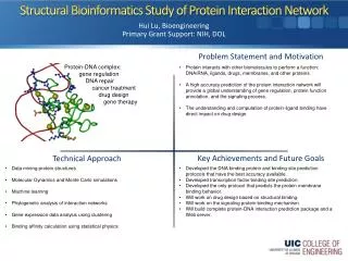
Protein structural element
Protein structural element. Yun-Ru (Ruby) Chen 陳韻如 Ph.D. The Genomics Research Center (office at 7th floor) yrchen@gate.sinica.edu.tw 2789-9930 ext 355. outline. Atom interaction and bonding Amino acid and peptide bond Secondary structure Tertiary structure Quantiary structure

Protein structural element
E N D
Presentation Transcript
Protein structural element Yun-Ru (Ruby) Chen 陳韻如 Ph.D. The Genomics Research Center (office at 7th floor) yrchen@gate.sinica.edu.tw 2789-9930 ext 355
outline • Atom interaction and bonding • Amino acid and peptide bond • Secondary structure • Tertiary structure • Quantiary structure • Function
Atom Interactions Energy Covalent interaction 300-400x Non-covalent interaction noncovalent interactions are 10-100 times weaker than covalent bonds.
Charge-charge interaction Coulomb's law defines the force between a pair of charges (q1 and q2) separated by a vacuum by a distance, r as F = k*(q1q2)/r2, where k is a constant. DE in vacuum=120kcal/M (very strong) In non-vacuum, dielectric medium F = k*(q1q2)/(D*r2) The dielectric constant arises from the fact that the dielectric medium shields the charges from each other. D water=79 dE in solution is lower because of hydration
Hydrogen bond • Hydrogen is shared between 2 electronegative atoms • Directional • Stronger than van der waal • Strength depends on donor and Acceptor electronegativity • (O>N>S)
Van der Waal Van der Waal radius of atoms
Peptide bond Carboxyl group(-COOH)=~2 Amide (-NH3+)=~9.6 Charged residues Acidic: Asp, D, pK1=~3.9, b-carboxyl. Glu, E, pKa=~4.3, g-carboxyl Basic: Lys, K, pK1=~10.5, b-carboxyl. Arg, R, pKa=~12.5, g-carboxyl His, H, pKa=~6, Hydroxyl residues Ser, pKa=~13.6 Thr, pKa=~13.6 Cys,pKa=~10.3 Protonation pKa<pH, deportonated pKa=pH, half-half pKa>pH, protonated pKa
Hydroxyl residues • Aliphatic • pKa=13.6
cis-trans Isomerization (trans:cis) • Non-proyl (1000:1) • X-proyl bond (4:1)
Disulfide bonds • Cysteine v.s. Cystine • Reducing agent DTT(dithiothreitol) TCEP (Tris[2-carboxyethyl] phosphine) Glutathione (reduced form vs. oxidized form (GSSG)) g-Glu-Cys-Gly
labeling • Amine-reacting group Reaction of a primary amine with an isothiocyanate Reaction of a primary amine with a succinimidyl ester or a tetrafluorophenyl (TFP) ester
Reaction of a primary amine with an STP ester Reaction of a primary amine with a sulfonyl chloride
Thiol group Reaction of a thiol with an alkyl halide Reaction of a thiol with a maleimide Reaction of a thiol with a symmetric disulfide (e.g., didansyl-L-cystine, D146).
Steric constrains dictate the possible types of secondary structure Ramachandran plot psi phi
Protein secondary structure Turn: beta turn, reverse turn, hairpin turn The simplest secondary structure element 3 or 4 aa involved
Helix • Alpha-helices are versatile cylindrical structures stabilized by a network of backbone hydrogen bonds • Helices can be right-handed (favored) or left-handed • 3.6aa per turn (a rotation of 100A) • 7aa for a helical wheel
Helical wheel Alpha-helices can be amphipathic with one polar and one non-polar face (favored helix-helix interaction) Lucine zipper
Special cases Collagen triple helix: proline found in left handed helices, three helices coil around each other Polyproline: when the peptide bonds are all trans it forms a left-handed helix with three residues per turn. Often serve as a docking sites for protein recognition modules such as SH3 domains in signal transduction pathways (exist in unfolded protein)
Beta sheets are extended structures that sometimes form barrels
Certain aa are more usually found in alpha helices, others in beta sheets • Long side chains are often found in helices • Side chain branched at b-carbon are often found in b stand • Proline and glycine are disfavored in helix and sheet • Predication is based on empirical rules (Chou-Fasman) • None is completely accurate
Condensed multiple secondary elements leads to tertiary structure (all alpha, all beta, mixed alpha/beta) V domain of IG light chain Triosephosphate isomerase Dihydrofolate reductase
Bound water In unfolded protein: backbond contacts with water In folded protein: water release from backbond contacts to bulk water, but water still interact with polar group on the surface either peptide bond and side-chains.
Hydrophobic effect • The tendency of nonpolar groups in water to self-associated and thereby minimize their contact surface are with the polar solvent • Exclusion of water • A driving force for folding
Reading Assignment • Chapter1 of Protein Structure and Function (or any other protein structure text book)