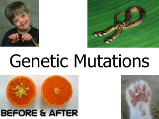
Types of Genetic Mutations
180 likes | 365 Vues
Types of Genetic Mutations. Main Types. Base Substitutions Gene Rearrangements Nondisjunction. Base Substitutions. Also known as “point” mutations, result when one base is substituted for another. Can be Silent Mutations Nonsense Mutations Missense Mutations. Silent Mutations.

Types of Genetic Mutations
E N D
Presentation Transcript
Main Types • Base Substitutions • Gene Rearrangements • Nondisjunction
Base Substitutions • Also known as “point” mutations, result when one base is substituted for another. • Can be • Silent Mutations • Nonsense Mutations • Missense Mutations
Silent Mutations • Cause no detectable change in the corresponding protein sequence • Most amino acids are encoded by several different codons so sometimes a change in the third base of a codon will have no affect on which amino acid in encoded. • For example, if the third base in the TCT codon for serine is changed to any one of the other three bases, serine will still be encoded. • Such mutations cannot be detected without sequencing the gene (or its mRNA).
Nonsense Mutation • Cause early termination of protein synthesis. • With a nonsense mutation, the new nucleotide changes a codon that specified an amino acid to one of the STOP codons (TAA, TAG, or TGA). Therefore, translation of the messenger RNA transcribed from this mutant gene will stop prematurely. The earlier in the gene that this occurs, the more truncated the protein product and the more likely that it will be unable to function. • Nonsense mutations occur in between 15% to 30% of all inherited diseases including cystic fibrosis, haemophilia, retinitis pigmentosa and duchenne muscular dystrophy.
Missense Mutations • Cause a different amino acid to be produced. • One example sickle-cell disease. The replacement of A by T at the 17th nucleotide of the gene for the beta chain of hemoglobin changes the codon GAG (for glutamic acid) to GTG (which encodes valine). Thus the 6th amino acid in the chain becomes valine instead of glutamic acid.
Here is a sampling of the more than 1000 different mutations that have been found in patients with cystic fibrosis. Each of these mutations occurs in a huge gene that encodes a protein (of 1480 amino acids) called the cystic fibrosis transmembrane conductance regulator (CFTR). The protein is responsible for transporting chloride ions through the plasma membrane. The gene encompasses over 6000 nucleotides spread over 27 exons on chromosome 7. • Defects in the protein cause the various symptoms of the disease. Unlike sickle-cell disease, then, no single mutation is responsible for all cases of cystic fibrosis. People with cystic fibrosis inherit two mutant genes, but the mutations need not be the same. • In some patients with cystic fibrosis, the substitution of a T for a C at nucleotide 1609 converted a glutamine codon (CAG) to a STOP codon (TAG). The protein produced by this patient had only the first 493 amino acids of the normal chain of 1480 and could not function.
Gene Rearrangments • Involve DNA sequences that have been modified, often by chemical and radioactive agents known as mutagens. • Can be • Deletions • Duplications • Inversions • Translocations
Deletions and Insertions • Result in the loss or gain of DNA or a gene. • Deletions can involve either the loss of a single base or the loss of a larger portions of DNA. The number can range from one to thousands. Insertions are similar… • Can have devastating consequences to the gene because of frameshift to the rest of the DNA sequence.
Several disorders in humans are caused by the inheritance of genes that have undergone insertions of a string of 3 or 4 nucleotides repeated over and over. • A locus on the human X chromosome contains such a stretch of nucleotides in which the triplet CGG is repeated (CGGCGGCGGCGG, etc.). The number of CGGs may be as few as 5 or as many as 50 without causing a harmful phenotype (these repeated nucleotides are in a noncoding region of the gene). Even 100 repeats usually cause no harm. However, these longer repeats have a tendency to grow longer still from one generation to the next (to as many as 4000 repeats). • This causes a constriction in the X chromosome, which makes it quite fragile. Males who inherit such a chromosome (only from their mothers, of course) show a number of harmful phenotypic effects including mental retardation. Females who inherit a fragile X (also from their mothers; males with the syndrome seldom become fathers) are only mildly affected.
Polyglutamine Diseases: In these disorders, the repeated trinucleotide is CAG, which adds a string of glutamines (Gln) to the encoded protein. These have beeen implicated in a number of central nervous system disorders including • Huntington's disease (where the protein called huntingtin carries the extra glutamines). The abnormal protein increases the level of the p53 protein in brain cells causing their death by apoptosis. • Some cases of Parkinson's disease where the extra glutamines are in the protein ataxin-2; • Some case of amyotrophic lateral sclerosis (ALS) — again where ataxin-2 is the culprit. (ALS is often called "Lou Gehrig's disease" after the baseball player who died from it.) • Muscular Dystrophy: Some forms of muscular dystrophy that appear in adults are caused by tri- or tetranucleotide, e.g. (CTG)n and (CCTG)n, repeats where n may run into the thousands. The huge RNA transcripts that result interfere with the alternative splicing of other transcripts in the nucleus.
Duplications • Can result in extra copies of genes, and are usually caused by unequal crossing over during meiosis or chromosome rearrangements.
Inversions • Occur when a section of a chromosome is broken away and then re-attached to the chromosome in an orientation opposite the original orientation (a 180 degree rotational shift). • May cause harmful effects if the inversion involves a gene or an important sequence involved in regulating gene expression.
Translocations • Occurs when a piece of one chromosome is transferred to a non-homologous chromosome . • They are often reciprocal, with the two chromosomes swapping segments with each other. • In most cases of chronic myelogenous leukaemia (CML), the leukaemic cells share a chromosomal abnormality known as Philadelphia chromosome. This abnormality is the result of a reciprocal translocation between chromosomes 9 and 22. An abnormal hybrid gene is created leading to the production of a novel protein that is not normally found in the cell. This protein prevents normal growth and development, leading to leukaemia.
Nondisjunction • Chromosome fail to separate properly during mitosis or meiosis. • Produces the wrong number of chromosomes in the cell. With the exception of Down’s Syndrome embryo will most likely not survive unless it occurs on the sex chromosomes. • Trisomy: extra chromosome, for example, Down’s Syndrome. • Monosomy: loss of chromosome, for example, Turner syndrome.
Humans inherit 3 x 10⁹ base pairs of DNA from each parent. Just considering single-base substitutions, this means that each cell has 6 billion (6 x 10⁹) different base pairs that can be the target of a substitution. • Single-base substitutions are most apt to occur when DNA is being copied; for eukaryotes that means during S phase of the cell cycle. • No process is 100% accurate. Even the most highly skilled typist will introduce errors when copying a manuscript. So it is with DNA replication. Like a conscientious typist, the cell does proofread the accuracy of its copy. But, even so, errors slip through. • It has been estimated that in humans and other mammals, uncorrected errors (= mutations) occur at the rate of about 1 in every 50 million (5 x 10⁷) nucleotides added to the chain. (Not bad — I wish that I could type so accurately.) But with 6 x 10⁹ base pairs in a human cell, that mean that each new cell contains some 120 new mutations. • Should we be worried? Probably not. Most (as much as 97%) of our DNA does not encode anything.
Although most mutations that change protein sequences are neutral or harmful, some mutations have a positive effect on an organism. In some cases, the mutation may enable the mutant organism to withstand particular environmental stresses better than wild-type organisms, or reproduce more quickly. In these cases a mutation will tend to become more common in a population through natural selection. • For example, a specific 32 base pair deletion in human CCR5 (CCR5-Δ32) confers HIV resistance to homozygotes and delays AIDS onset in heterozygotes. The CCR5 mutation is more common in those of European descent. One possible explanation of the etiology of the relatively high frequency of CCR5-Δ32 in the European population is that it conferred resistance to the bubonic plague in mid-14th century Europe. People with this mutation were more likely to survive infection; thus its frequency in the population increased. This theory could explain why this mutation is not found in southern Africa, where the bubonic plague never reached. A newer theory suggests that the selective pressure on the CCR5 Delta 32 mutation was caused by smallpox instead of the bubonic plague. • Another example, is Sickle cell disease which is a blood disorder in which the body produces an abnormal type of the oxygen-carrying substance hemoglobin in the red blood cells. One-third of all indigenous inhabitants of Sub-Saharan Africa carry the gene, because in areas where malaria is common, there is a survival value in carrying only a single sickle-cell gene (sickle cell trait). Those with only one of the two alleles of the sickle-cell disease are more resistant to malaria, since the infestation of the malaria plasmodium is halted by the sickling of the cells which it infests.
All types of mutations, if not repaired, will be kept in subsequent rounds of replication. Mutations in somatic cells may damage the cell, make it cancerous or kill it. Mutations in a germ cell (cells that give rise to gametes) will be passed down to the next generation.