Download
1 / 9
90 likes | 130 Vues
A 50-year-old man underwent sigmoidoscopy for GI pathology evaluation. The findings revealed a benign neoplasm known as a Villous Adenoma characterized by finger-like mucosal projections. The epithelium lining the villi showed dysplastic changes with enlarged, hyperchromatic cells. The adjacent mucosa appeared normal.

E N D
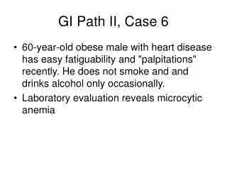
GI Pathology II, Case 5 • 50-year-old man has sigmoidoscopy to evaluate for GI pathology
A Villous Adenoma B Tubular Adenoma
Describe the histologic findings. Diagnosis?
Villous adenoma Normal colonic mucosa Villous Adenoma A low power view of the colonic mucosa revelas a benign neoplasm composed of villi or "finger-like" mucosal projections (*). The adjacent mucosa contains normal glands.
A section of the colon reveals a broad based polyp which for the most part forms villi (>50%)
Villi are lined by an epithelium which is dysplastic, with cells containing enlarged, hyperchromatic and pseudostratified epithelium