Download
1 / 20
200 likes | 223 Vues
PART 1. The Special Senses. The Special Senses. Taste, smell, sight, hearing, and balance Touch – a large group of general senses Special sensory receptors Localized – confined to the head region Receptors are not free endings of sensory neurons Special receptor cells.

E N D
PART 1 The Special Senses
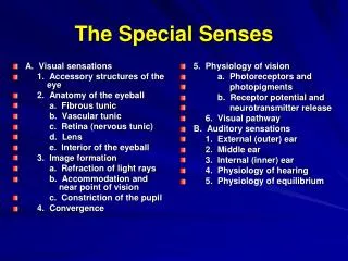
The Special Senses • Taste, smell, sight, hearing, and balance • Touch – a large group of general senses • Special sensory receptors • Localized – confined to the head region • Receptors are not free endings of sensory neurons • Special receptor cells
The Chemical Senses: Taste and Smell • Taste – gustation • Smell – olfaction • Receptors – classified as chemoreceptors • Respond to chemicals
Taste – Gustation • Taste receptors • Occur in taste buds • Most are found on the surface of the tongue • Located within tongue papillae • Two types of papillae (with taste buds) • Fungiform papillae • Circumvallate papillae • NOTE: Filiform papillae do NOT contain taste buds • Filiform papillae help you appreciate texture of food
Taste Buds • Collection of 50 –100 epithelial cells • Contain three major cell types (similar in all special senses) • Supporting cells • Gustatory cells • Basal cells • Contain long microvilli – extend through a taste pore
Taste Buds Figure 16.1a, b
Taste Sensation and the Gustatory Pathway • Four basic qualities of taste • Sweet, sour, salty, and bitter • A fifth taste – umami, “deliciousness” • No structural difference among taste buds
Gustatory Pathway • Taste information reaches the cerebral cortex • Primarily through the facial (VII) and glossopharyngeal (IX) nerves • Some taste information through the vagus nerve (X) • Sensory neurons synapse in the medulla • Located in the solitary nucleus
Gustatory Pathway from Taste Buds Figure 16.2
Smell (Olfaction) • Receptors are part of the olfactory epithelium • Olfactory epithelium composed of • Cell bodies of olfactory receptor cells • Supporting cells – columnar cells • Basal cells – form new olfactory receptor cells
Smell (Olfaction) • Axons of olfactory epithelium • Gather into bundles – filaments of the olfactory nerve • Pass through the cribriform plate of the ethmoid bone • Attach to the olfactory bulbs
Olfactory Receptors Figure 16.3a, b
Disorders of the Chemical Senses • Anosmia – absence of the sense of smell • Due to injury, colds, allergies, or zinc deficiency • Uncinate fits – distortion of smells or olfactory hallucinations • Often result from irritation of olfactory pathways • After brain surgery or head trauma
Embryonic Development of the Chemical Senses • Development of olfactory epithelium and taste buds • Olfactory epithelium – derives from olfactory placodes • Taste buds develop upon stimulation by gustatory nerves
The Eye and Vision • Visual organ – the eye • 70% of all sensory receptors are in the eyes • 40% of the cerebral cortex is involved in processing visual information • Anterior one-sixth of the eye’s surface is visible
Accessory Structures of the Eye • Eyebrows – coarse hairs on the superciliary arches • Eyelids (palpebrae) – separated by the palpebral fissure • Meet at the medial and lateral angles (canthi) • Lacrimal caruncle – reddish elevation at the medial canthus • Tarsal plates – connective tissue within the eyelids • Tarsal glands – modified sebaceous glands
Accessory Structures of the Eye • Conjunctiva – transparent mucous membrane • Palpebral conjunctiva • Bulbar conjunctiva • Conjunctival sac Figure 16.5a
Accessory Structures of the Eye • Lacrimal apparatus – keeps the surface of the eye moist • Lacrimal gland – produces lacrimal fluid • Lacrimal sac – fluid empties into nasal cavity Figure 16.5b
Extrinsic Eye Muscles • Six muscles that control movement of the eye • Originate in the walls of the orbit • Insert on outer surface of the eyeball • Annular ring – origin of the four rectus muscles PLAY Accessory Eye Structures
Extrinsic Eye Muscles Figure 16.6a, b