Download
1 / 40
400 likes | 519 Vues
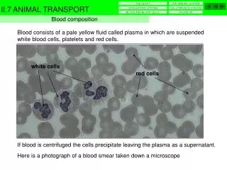
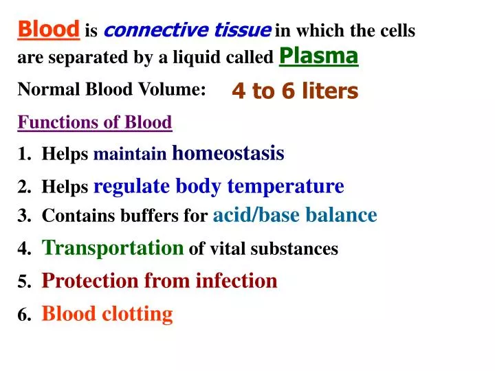
Blood is connective tissue in which the cells are separated by a liquid called Plasma. Normal Blood Volume:. 4 to 6 liters. Functions of Blood. 1. Helps maintain homeostasis. 2. Helps regulate body temperature. 3. Contains buffers for acid/base balance.

E N D
Bloodis connective tissue in which the cells are separated by a liquid called Plasma Normal Blood Volume: 4 to 6 liters Functions of Blood 1. Helps maintain homeostasis 2. Helps regulate body temperature 3. Contains buffers for acid/base balance 4. Transportation of vital substances 5. Protection from infection 6. Blood clotting
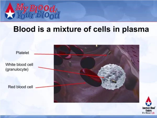
Formed Elements of Blood 1. Red Blood Cells (RBC's) -- make up about 45% of our blood volume -- old RBC removed from the blood by the liver & spleen -- Erythrocytes -- live for 120 days or 4 months -- normal count: 4.2 to 6.2 million/mm3
2. White Blood Cells (WBC's) 3. Platelets -- Leukocytes -- normal count: 5,000 to 10,000/mm3 -- Thrombocytes -- normal count: 150,000 to 300,000 /mm3 -- essential for blood clotting -- live only 10 days -- have no nucleus -- considered a cell fragment
Cells that are found in the Red Bone Marrow are: -- which give rise to all your blood STEM CELLS cells -- their large number & unique shape increases their total surface area equal to a football field -- RBC's transport oxygen, carbon dioxide, & hydrogen ions (RBC, WBC, & Platelets) RBC's have a thin center & thicker edges A red pigment located on RBC's that carries Oxygen is called: Hemoglobin -- when combined with O2 it's called: Oxyhemoglobin -- Oxyhemoglobin is bright red -- hemoglobin without oxygen is dark purple
RBC's WBC's Plasma LAB TESTS
DIFFERENTIAL • ----Looks at the number of each type of • WBC and it’s shape. ----Gives specific information about a patient’s immune system.
The nuclei in mature Neutrophils are divided into segments, so are calledSEGS. Fast-actingNeutrophils are the first line of defense against bacteria. Survive only 4 to 10 hours. Eosinophilsare weak phagocytes, but good at detoxifying allergens. Also, defend against parasites. Basophils secrete Histamine, Heparin, and Serotonin. They are involved in systemic hypersensitivity reactions. Immature Neutrophils have unsegmented nuclei that look like bands, so are calledBANDS RED BLOOD CELLS Monocytesare phagocystic and produce substances that mark invading organism for destruction by lymphocytes. Slower than Neutrophil, but last longer. PLATELETS STEM CELL Lymphocytesinclude T-cells, which turn immunity on or off, and B-cells, which produce antibodies.
Blood Formed Elements Plasma H2O Water Salts Dissolved gases Hormones Glucose Wastes Plasma Proteins Red Blood Cells Platelets White Blood cells Albumin Fibrinogen Granulocytes Neutrophils Eosinophils Basophils Agranulocytes Monocytes Lymphocytes Globulins Prothrombin Complements enzymes that help antibodies fight infections
Steps in Blood Clotting • Platelet Factors combine with Prothrombin (an inactive enzyme) and Calcium to form Thrombin (an active enzyme) -- Vitamin K is needed to stimulate the liver to produce more Prothrombin • Thrombin reacts with Fibrinogen to form a fibrous gel called Fibrin (a clot) • Injury to blood vessel • Platelets break up & release Platelet factors
Trauma Activated factor XII Factor XII Activated factor XI Factor XI Factor IX Activated factor IX Ca+ Factor VIII Factor X Activated factor X Prothrombin activator Ca+ Thrombin Prothrombin Fibrinogen Fibrin Ca+ BLOOD CLOTTING Platelet Platelet plug Fibrin
For normal clotting, we need: Platelets & Platelet Factors Calcium Blood Proteins Prothrombin & Fibrinogen Vitamin K A clot that remains stationary in the blood vessel is called: a Thrombus A dislodged blood clot that moves in the blood is called an: Embolus Partially blocked coronary arteries will cause: Ischemic Heart Disease This can cause pain during exercise and stress called: Angina Pectoris
Myocardial Infarction or a M.I. -- still contain Antibodies, so can be used to treat patient who have a need for a specific antibody If the coronary artery is totally blocked, the heart tissue will die. This is called a: Serum -- Blood Plasma minus its clotting factors which are: Prothrombin Fibrinogen
-- A Substance that can stimulate the body to make Antibodies -- is a Substance that reacts with theAntigen that stimulated its Formation TYPE A ANTIGEN (located on the RBC) ANTI-B ANTIBODIES (located in the Plasma) BLOOD TYPES ANTIGEN ANTIBODY -- causes the Antigen to agglutinate(clump) TYPE A TYPE B TYPE B ANTIGEN ANTI-A ANTIBODIES
NO ANTIGEN TYPE A ANTIGEN & (located on the RBC) TYPE B ANTIGEN ANTI-A ANTIBODIES O & O ANTI-B ANTIBODIES (located in the Plasma) B A B A AB AB BLOOD TYPES TYPE O TYPE AB Universal Donor Universal Recipient NO ANTIBODIES
TYPE Rh ANTIGEN (located on the RBC) NO ANTIBODIES ANTI-Rh ANTIBODIES will be produced with (located in the Plasma) exposure BLOOD TYPES TYPE Rh - Positive TYPE Rh - Negative NO ANTIGEN Negative can give to Negative and to Positive Positive can only give to Positive
Plasma never naturally contains Anti-Rh Antibodies -- at delivery a little of baby's blood mixes with Mom's blood when placenta separates antibodies -- not a problem with first pregnancy, but with each subsequent pregnancy, Mom makes more -- eventually she has enough antibodies to cross the placental barrier and attack baby's blood -- give Rho Gam to prevent production of antibodies -- causes a problem when Mom is Rh-negative. and has a babythat is Rh-positive -- this stimulates Mom's blood to make antibodies -- a condition called Erythroblastosis Fetalis
Membranes of the heart Epicardium or Visceral Pericardium Myocardium Endocardium Covering of the heart is called: Pericardium Membrane that covers surface of heart: Middle layer is major portion of the heart, is largely cardiac muscle, & is called: Membrane that lines the heart chambers:
Aorta HEART Superior vena cava Right Left Right Atrium Pulmonary Atrium veins Right Left Inferior vena cava Ventricle Ventricle Tricuspid valve Bicuspid or Pulmonary Mitral Valve arteries Aortic Cordae Pulmonary valve valve tendineae Papillary Apex Septum muscle
PULMONARY CIRCULATION SYSTEMIC CIRCULATION Blood enters the RIGHT ATRIUM from the SUPERIOR & INFERIOR VENA CAVAS LUNGS RIGHT & LEFT PULMONARY VEINS RIGHT ATRIUM LEFT ATRIUM TRICUSPID VALVE BICUSPID OR MITRAL VALVE RIGHT VENTRICLE LEFT VENTRICLE PULMONARY VALVE AORTIC VALVE PULMONARY TRUNK AORTA BODY Rt.. & Lt.PULMONARY ARTERIES LUNGS
Heart Sounds Pulmonary Valve Aortic Valve Tricuspid Mitral Valve Valve When you listen to the heart, the sounds you hear are the valves closing The first sound (Lup) is the: closing The second sound (Dup) is the: closing
AV Bundle or Bundle of HIS Lt. atrium SA node Rt. Atrium AV node Purkinje Fibers Rt. Ventricle Lt. Ventricle Lt. Bundle Branch Rt. Bundle Branch Cardiac Conduction Atriums contract Ventricles contract
-- a graph of the electrical activity of the heart -- the P-wave signifies the atriums contracting -- the QRS-wave signifies the ventricles contracting QRS -- the T-wave signifies the relaxing of the ventricles T-wave P-wave EKG
Terms Heart Beat (HR) -- heart rate greater than 100 beats/min. -- the volume of blood ejected Stroke Volume from the ventricles during each beat (SV) Cardiac Output (CO) -- volume of blood pumped by one ventricle per minute Systole Diastole -- Number of beats of the heart per min. (Average is 70 beats per minute) Bradycardia -- heart rate less than 60beats/min. Tachycardia (Average = 70 cc/beat.) (Average = 5 Liters) Cardiac output=(HR x SV)70 x 70=4900 cc ~ 5L/min. Cardiac Cycle -- the complete heart beat
left coronary artery -- blood flows into the heart by way of the right & left coronary arteries -- coronary arteries are the Aorta's first branches -- this way the blood with the highest % of O2 is delivered to the heart muscle right coronary artery -- the coronary arteries fill when the ventricles are relaxed --veins are removed from other areas of the body & used to bypass the blockage in the coronary artery Coronary Circulation Coronary Artery Bypass Surgery
Artery -- take blood away from the Arteriole heart -- contain large amount of elastic fibers to accommodateincrease in blood volume with each heart beat Capillary -- blood flow is fastest in the arteries Types of Blood Vessels -- are small arteries that carry blood to the capillaries -- under control of the ANS (Sympathetic) -- whether dilated or constricted, affects blood pressure
-- exchange of nutrients and waste molecules takes place here -- O2 & glucose diffuse out & CO2 diffuses in Venule Capillary -- wall are thinner & less CO2 elastic than arteries Vein O2 -- small vessels that drain blood from the capillaries & then join together to form a vein -- blood flow is the slowesthere because: -- takes blood to the heart -- contains valves to prevent back flow
Rt. carotid Lt. common carotid Rt. subclavian Lt. subclavian Brachiocephalic -- supplies lt. side of head Branches of the Aorta -- forms the right carotid & rt. subclavian -- supplies the rt. side of the head & rt. arm -- supplies left arm
Common Iliac Arteries Femoral Popliteal Posterior tibial Pedal Other Systemic Arteries
-- refers to blood flow Inferior vena cava through the liver Hepatic Vein -- digestive organs send their blood to the liver by way of the Hepatic Portal Vein -- Blood leaves the liver by the Hepatic veins to the Inferior Vena Cava Hepatic Portal Circulation -- also called Portal Circulation -- this detour serves 2 functions: 1. remove excess glucose for storage as glycogen 2. remove & detoxify any poisonous substances
Blood Pressure -- liquids can only flow from an area of higher -- blood does not circulate pressure to an area of lower. if not present BP in Vena Cava is 0 BP in Aorta is 100 mm Hg -- or thedifference between the beginning& the endof a vessel fastest? slowest? -- the pressure or push of blood Capillaries Arteries -- exists in all blood vessels Blood Pressure Gradient -- it is the difference between 2blood pressures -- pressure drops throughout the vessel's length
Systolic Blood Pressure -- pressure in an artery when left ventricle is contracting -- pressure in an artery when left ventricle is 120 resting = -80 -- Difference between the Systolic & the Diastolic -- expressive of the health of the heart & tone of the arteries blood pressure Diastolic Blood Pressure Textbook BP: 120/80 40 Pulse Pressure -- over 50points or under 30is considered abnormal (hypertension or ICP) (shock)
-- vessel expands & then Carotid Dorsalis returns to normal pedis Temporal Apical -- place fingertips over artery, & press it over a Brachial bone or other firm Radial surface -- Provides information Popliteal about the heart beat: Femoral Posterior tibial Pulse -- surge of blood entering the artery 1. Rate 2. Strength 3. Rhythm
-- pressure in the arterioles forces fluid into Lymph the interstitial spaces Capillary Tissue Fluid Cells Arteriole -- most of this fluid will be returned to the venules -- what doesn't, enter the lymphatic capillaries Venule Blood Capillary -- lymph capillaries are similar to veins because they contain valves & the fluid is moved by muscle contraction Lymph -- specialized fluid formed in the tissue spaces -- this fluid is called: Interstitial Fluid -- this fluid is now called: Lymph
Thoracic Duct & Right Lymphatic Duct -- which then returns the lymph fluid to the venous circulation Function of the Lymphatic System 2. Transport fluids to the blood stream -- lymph veins empty into: 1. Produce Lymphocytes 3. Absorb fat molecules
-- fluid enters the node by way of an afferent vessel -- important for the maturation & maintenance of the immune system & especially the T-Cells Organs of the Lymphatic System Lymph Node -- clustered along the lymphatic vessels -- Function 1. Defense -- filter the lymph fluid -- fluid leaves by way of an efferent vessel 2. White Blood Cell Formation Thymus Gland -- Produces: Thymosin -- decreases in size with age
2. Destroys worn out RBC's & salvages the iron in hemoglobin 3. Serves as a reservoir for blood that can be returned to the circulatory system when needed Spleen -- largest lymphoid organ in the body -- located upper left quadrant of the abdomen -- protected by the ribs, but can be injured -- Functions: 1. Filters the blood -- what other organ does this? Liver -- it stores up to 1 pint of blood Lymph nodes clean lymph fluid & the spleen cleans the blood
-- composed of lymphoid tissue located in the mouth & throat Palatine tonsils Pharyngeal tonsils Lingual tonsils Tonsils 1 . "tonsils" 2. "adenoids" 3. near the base of the tongue -- serves as the first line of defense from the exterior -- removal of the palatine tonsils is called: Tonsillectomy -- removal of the pharyngeal tonsils is called: Adenoidectomy
1. Bacteria 2. Foreign tissue cells 3. Cancerous cells ----Originates from the Stem Cells in our Bone Marrow ----Goes through 2 stages of development ----Protects us from:
2. As with B-Cells, 2nd stage begins with contact with aspecific antigen. ----First stage of development occurs in the Thymus gland. 1. Ends up in the lymph nodes. 3. Functions in Cell-mediated Immunity.
Lymphocytes B-CELLS T-CELLS Lymphoid Tissue Plasma Cells Memory Cells Sensitized T-Cells Suppressor Killer Helper Memory Antibodies T-Cells T-Cells T-Cells T-Cells (Stimulates other immune cells including B & Killer T-Cells) (Stops the immune response)