Analysis of NSMCE2, TRIBI1, and MYC Rearrangements in Patient 1
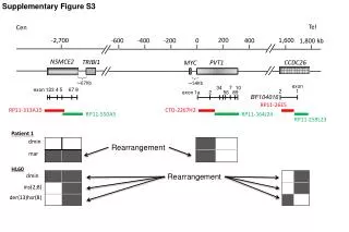
This study investigates the genomic rearrangements involving NSMCE2, TRIBI1, PVT1, CCDC26, and MYC in Patient 1. Using data from Supplementary Figure S3, we explore the structural variations represented by markers and exon distributions. Notably, the analysis includes significant alterations such as ins(2;8) and der(13)hSR(8). The involvement of specific exons and their relative positions across a span of approximately 67Kb to 54Kb from the telomeres to centromeres sheds light on potential pathways influencing tumorigenesis.

Analysis of NSMCE2, TRIBI1, and MYC Rearrangements in Patient 1
E N D
Presentation Transcript
Supplementary Figure S3 200 -2,700 1,600 -600 -400 -200 0 400 1,800 kb NSMCE2 TRIBI1 PVT1 CCDC26 MYC ~67Kb ~54Kb Tel Cen exon exon 123 4 5 67 8 2 1 exon 1a 2 56 BF104016 10 34 7 RP11-26E5 89 RP11-313A10 CTD-2267H2 RP11-550A5 RP11-164J24 RP11-259L23 Patient 1 dmin Rearrangement mar HL60 dmin Rearrangement ins(2;8) der(13)hsr(8)