Download
1 / 33
330 likes | 339 Vues
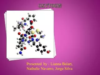
Role of oxytocin in energy metabolism. Peptides 45 (2013) 9–14. Valéria Ernestânia Chaves Federal University of São João Del Rei Brazil. Oxytocin. First peptide hormone whose structure was determined and the first to be chemically synthesized in its biologically active form.

E N D
Role of oxytocin in energy metabolism Peptides 45 (2013) 9–14. Valéria Ernestânia Chaves Federal University of São João Del Rei Brazil
Oxytocin First peptide hormone whose structure was determined and the first to be chemically synthesized in its biologically active form. Du Vigneaud V, Ressler C, Trippett S. The sequence of amino acids in oxytocin, with a proposal for the structure of oxytocin. J Biol Chem 1953; 205:949–57.
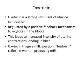
Functions of Oxytocin • OT affects: • Myometrium, stimulating uterine contraction and • Myoepithelial cells of the mammary gland, stimulating the milk ejection. Dale HH. On some physiological actions of ergot. J Physiol (Lond) 1906; 34:163–206. Ott J, Scott JC. The galactogogue action of the thymus and corpus luteum. Proc Soc Exp Biol1910; 8:49.
Functions of Oxytocin OT exerts several central influences, from modulating neuroendocrine reflexes to establishing the complex social behaviors related to reproduction and the care of offspring as well as learning and memory. Feldman R, Weller A, Zagoory-Sharon O, Levine A. Evidence for a neuroen-docrinologicalfoundationofhumanaffiliation: plasma oxytocinlevelsacrosspregnancyandthe post partumperiodpredictmother-infantbonding. PsycholSci 2007;18:965–70. Guastella AJ, Mitchell PB, Dadds MR. Oxytocin increases gaze to the eye regionof human faces. Biol Psychiatry 2008;63:3–5. Insel TR, Young L, Wang Z. Central oxytocin and reproductive behaviours. RevReprod 1997;2:28–37. Kosfeld M, Heinrichs M, Zak PJ, Fischbacher U, Fehr E. Oxytocinincreasestrustinhumans. Nature 2005;435:673–6. Ross HE, Young LJ. Oxytocin and the neural mechanisms regulating social cognition and affiliative behavior. Front Neuroendocrinol 2009;30:534–47.
Functions of Oxytocin The physiological importance of OT in metabolic homeostasis has also been reported. Camerino C. Lowsympathetic tone andobesephenotype in oxytocin-deficientmice. Obesity 2009;7:980–4. Takayanagy Y, Kasahara Y, Onaka T, Takahashi N, Kawada T, Nishimori K.Oxytocin receptor-deficient mice developed late-onset obesity. Neuroreport2008;19:951–5. Zhang G, Cai D. Circadian intervention of obesity development via resting-stagefeeding manipulation or oxytocin treatment. Am J Physiol Endocrinol Metab2011;301:E1004–12.
Mice deficient in OT receptor develop late-onset obesity Takayanagy Y, Kasahara Y, Onaka T, Takahashi N, Kawada T, Nishimori K.Oxytocin receptor-deficient mice developed late-onset obesity. Neuroreport 2008; 19:951–5.
Mice deficient in OT receptor develop late-onset obesity Takayanagy Y, Kasahara Y, Onaka T, Takahashi N, Kawada T, Nishimori K.Oxytocin receptor-deficient mice developed late-onset obesity. Neuroreport 2008; 19:951–5.
Mice deficient in OT receptor develop late-onset obesity Takayanagy Y, Kasahara Y, Onaka T, Takahashi N, Kawada T, Nishimori K.Oxytocin receptor-deficient mice developed late-onset obesity. Neuroreport 2008; 19:951–5.
Mice deficient in OT receptor develop late-onset obesity Takayanagy Y, Kasahara Y, Onaka T, Takahashi N, Kawada T, Nishimori K.Oxytocin receptor-deficient mice developed late-onset obesity. Neuroreport 2008; 19:951–5.
Mice deficient in OT receptor develop late-onset obesity Takayanagy Y, Kasahara Y, Onaka T, Takahashi N, Kawada T, Nishimori K.Oxytocin receptor-deficient mice developed late-onset obesity. Neuroreport 2008; 19:951–5.
Mice deficient in OT receptor develop late-onset obesity despite normal food intake and motor activity Takayanagy Y, Kasahara Y, Onaka T, Takahashi N, Kawada T, Nishimori K. Oxytocin receptor-deficient mice developed late-onset obesity. Neuroreport 2008;19:951–5.
Mice deficient in OT also increase the body weight accompanied by a 40% increase in abdominal fat pads Figure 1 Body weight and fat mass. Body weight curves of Oxt−/− (n = 10) and Oxt+/+ (n = 10) male and female mice. No body weight difference between control and knockout mice until 2nd month of age has been observed. Differences in the body weight between Oxt−/− and wild-type mice were observed at 3rd month of age. No differences between gender were observed within Oxt−/− or Oxt+/+ mice. *Significantly different with respect to Oxt+/+ with P < 0.005 (a). Weight of abdominal fat pad in 6 months old Oxt−/− and Oxt+/+ males and females mice. *Significantly different with respect to Oxt+/+ with P < 0.05 (b). Camerino C. Low sympathetic tone and obese phenotype in oxytocin-deficient mice. Obesity 2009;7:980–4.
Mice deficient in OT develop obesity with normal food intake and increase in leptin levels 4th month of age Figure 2 Food intake and leptin plasma levels. Food intake per day in Oxt+/+ (n = 5) and Oxt−/− (n = 5) male mice (a). The numbers indicate the four consecutive days of measurements. The histogram referred to a food measurement performed at 4th month of age. However, the same results have been obtained at each time. Plasma leptin concentration in male Oxt+/+ (n = 8) and Oxt−/− (n = 8) mice and in female Oxt+/+ (n = 5) and Oxt−/− (n = 5) mice at 6 months of age. *Significantly different with respect to Oxt+/+ with P < 0.05 (c). Camerino C. Low sympathetic tone and obese phenotype in oxytocin-deficient mice. Obesity 2009;7:980–4.
Central OT infusion causes a lower body weight gain in diet-induced obese rats. > 50% Figure 1. Central OT infusion causes body weight loss independently from changes in food intake. The measurements were performed over a 14-day experimental period (weeks 5 through 7 of a high fat diet): (A) Cumulative body weight changes; (B) cumulative food intake. Filled bars: i.c.v. saline–infused controls; open bars: i.c.v. OT-infused rats (1.6 nmol/d). Values are mean 6 SEM of 6 to 7 rats/group. *P,0.05 compared to controls. Deblon N, Veyrat-Durebex C, Bourgoin L, Caillon A, Bussier AL, Petrosino S, et al. Mechanisms of the anti-obesity effects of oxytocin in diet-induced obese rats. PLoS ONE 2011;6(9):e25565.
OT modulates the peripheral metabolism LipidMetabolism
The continuous ICV infusion of OT (1.6 nmol/day for 14 days) caused a body weight loss independently of the changes in food intake and induced an increase in the plasma glycerol levels, which was accompanied by a decrease in the plasma triacylglycerol levels without changes in the plasma insulin, leptin, and glucose levels Table. Effects of i.c.v. oxytocin (1.6 nmol/d) infusion on plasma glucose, insulin, leptin, FFA, glycerol, TG, oleoylethanolamide (OEA), palmitoylethanolamide (PEA), anandamide (AEA) and 2-arachidonoylglycerol (2-AG) levels. Deblon N, Veyrat-Durebex C, Bourgoin L, Caillon A, Bussier AL, Petrosino S, et al. Mechanisms of the anti-obesity effects of oxytocin in diet-induced obese rats. PLoS ONE 2011;6(9):e25565.
The continuous ICV infusion of OT (1.6 nmol/day for 14 days) also induced an increase in the expression and content of hormone-sensitive lipase in adipose tissue, suggesting higher lipolytic activity in this tissue. Figure. Central OT infusion stimulates lipid metabolism. The following analyses were performed on epididymal white adipose tissue (eWAT) of i.c.v. saline–infused controls (filled bars) and i.c.v. OT-infused rats (1.6 nmol/d; open bars): (A) mRNA expression of enzymes related to lipid metabolism; and (B) Western blot analysis of HSL standardized to actin expression; Values are mean ± SEM of 6 to 7 rats/group. *P>0.05, **P>0.01 compared to controls. Deblon N, Veyrat-Durebex C, Bourgoin L, Caillon A, Bussier AL, Petrosino S, et al. Mechanisms of the anti-obesity effects of oxytocin in diet-induced obese rats. PLoS ONE 2011;6(9):e25565.
Central OT infusion induces hypothalamic OT synthesis and release into the bloodstream Figure 4. Central OT infusioninduceshypothalamic OT synthesis and release into the bloodstream. The following parameters were measured at the end of 14-day treatments with two doses of i.c.v. OT infusion: (A) Oxytocin expression (Oxt) in rat hypothalamus; (B) plasma OT levels in saline–infused controls (filled bars) and OT-infused rats (1.6 nmol/d, open bars). Values are mean 6 SEM of 6 to 7 rats/group. *P<0.05 compared to controls. Deblon N, Veyrat-Durebex C, Bourgoin L, Caillon A, Bussier AL, Petrosino S, et al. Mechanisms of the anti-obesity effects of oxytocin in diet-induced obese rats. PLoS ONE 2011;6(9):e25565.
Incubating epididymal adipose tissue with OT (10 nM for 4 h) increased the glycerol content in the incubation medium. Figure 5. OT directly affects lipid metabolism (B–C) Epididymal fat pads from lean Wistar rats were incubated at 37ºC in the presence of Krebs-Ringer-Hepes buffer containing 2% FA-free BSA and 0.1% glucose. After 4 h of incubation in the presence of either saline or OT (10 nM), the amount of (B) glycerol and (C) free fatty acid released in the medium was measured. Values are mean 6 SEM of three independent experiments. Deblon N, Veyrat-Durebex C, Bourgoin L, Caillon A, Bussier AL, Petrosino S, et al. Mechanisms of the anti-obesity effects of oxytocin in diet-induced obese rats. PLoS ONE 2011;6(9):e25565.
A peripheral and continuous OT treatment (3.6 mg/100 g−1 body weight per day for 14 days) of Wistar rats fed with a commercial diet decreased the diameter of the adipocytes without changing adipose tissue mass. Eckertova M, Ondrejcakova M, Krskova K, Zorad S, Jezova D. Subchronic treat-ment of rats with oxytocin results in improved adipocyte differentiation andincreased gene expression of factors involved in adipogenesis. Br J Pharmacol 2011;162:452–63.
OT treatment increased number of small adipocytes and the expression of the PPAR-gamma gene, an important transcriptional factor involved in adipogenesis, in epididimal adipose tissue. Eckertova M, Ondrejcakova M, Krskova K, Zorad S, Jezova D. Subchronic treat-ment of rats with oxytocin results in improved adipocyte differentiation andincreased gene expression of factors involved in adipogenesis. Br J Pharmacol 2011;162:452–63.
Peroxisome proliferator activated receptor (PPAR)-alpha may mediate the body weight gain control, because a peripheral administration of OT (50 nmol for 3 days) did not affect the body weight gain in PPAR-alpha knockout mice but did decrease this parameter in wild-type mice. Figure 7. PPAR-alpha mediates peripheral OT effects. (A) Cumulative body weight gain after 3 days of s.c. saline or OT treatment in PPARalpha KO and wild-type (WT) mice. Values are mean ± SEM of 5 animals/group. *P < 0.05 compared to controls. Deblon N, Veyrat-Durebex C, Bourgoin L, Caillon A, Bussier AL, Petrosino S, et al. Mechanisms of the anti-obesity effects of oxytocin in diet-induced obese rats. PLoS ONE 2011;6(9):e25565.
The peripheral OT treatment of obese mice fed a high-fat diet induced a decrease in the respiratory quotient, specifically during the light phase, without significantly altering the energy expenditure or the locomotor activity, suggesting that OT promotes the use of fat as an energy substrate. Figure 4. Chronic Oxt infusion promotes use of fat. (A, B) Effect of chronic Oxt infusion on (A) time course of respiratory quotient (RQ) (B) average RQ in the light and dark phases (C) time course of energy expenditure (EE) (D) average values for the light and dark phases (E) time course of cumulative locomotor activity every 0.5 hr (F) average values in the light and dark phases. n = 5 in each group. *p < 0.05, **p < 0.01. Maejima Y, Iwasaki Y, Yamahara Y, Kodaira M, Sedbazar U, Yada T. Peripheral oxytocin treatment ameliorates obesity by reducing food intake and visceral fat mass. Aging (Albany, NY) 2011;3:1169–77.
OT modulates the peripheral metabolism Glucose Metabolism
The presence of oxytocin in the incubation medium (1 mol/L for 20 min) induced an increase in the rate of glucose 1-14C oxidation to CO2 in adipocytes . 40% Fain JN, Gokmen-Polar Y, Bahouth SW. Wortmannin converts insulin but not oxytocin from an antilipolytic to a lipolytic agent in the presence of forskolin. Metabolism 1997;46:62–6.
OT was shown to stimulate the glucose oxidation and lipogenesis in adipocytes Hanif K, Goren HJ, Hollenberg MD, Lederis K. Oxytocin action: mechanisms forinsulin like activity in isolated rat adipocytes. Mol Pharmacol 1982;22:381–8.
Whether infused centrally or peripherally, chronic OT infusion improved the insulin sensitivity of diet-induced obese rats . Figure. Central OT infusion protects against high fat diet-induced insulin resistance. I.c.v. saline- (black circles) or OT- (1.6 nmol/d; white diamonds) infused rats received: glucose tolerance tests (1.5 g/kg) before (black triangles, dashed line; 3 weeks of HFD; n = 16 rats) or after infusions (7 weeks of HFD; 14-day i.c.v. infusions; n = 6 for each treatment group): (A) delta glucose and (B) delta insulin; One-way ANOVA: *P<0.05 compared to black triangles; {P,0.05 compared to black circles. Deblon N, Veyrat-Durebex C, Bourgoin L, Caillon A, Bussier AL, Petrosino S, et al. Mechanisms of the anti-obesity effects of oxytocin in diet-induced obese rats. PLoS ONE 2011;6(9):e25565.
Mice deficient in OT have an insulin-resistant state and a lower capability to counteract the glycemia increase following a glucose bolus Figure. Glucose metabolism. Insulin tolerance test (ITT) (a). Representative plots of glucose disposal curves for each experimental condition. (b). Representative plots of glucose disposal curves for each experimental condition.The data are the mean ± ES of blood glucose values from three wild-type and three Oxt−/− mice normalized on the starting glucose values at time 0 for each mice. *Significantly different with respect to Oxt+/+ with P < 0.05. Camerino C. Low sympathetic tone and obese phenotype in oxytocin-deficient mice. Obesity 2009;7:980–4.
Peripheral OT increased the GLUT-4 (an insulin-dependent glucose transporter) expression in the epididymal adipose tissue . Eckertova M, Ondrejcakova M, Krskova K, Zorad S, Jezova D. Subchronic treat-ment of rats with oxytocin results in improved adipocyte differentiation andincreased gene expression of factors involved in adipogenesis. Br J Pharmacol 2011;162:452–63.
OT directly stimulated the insulin release from mice islets via phosphoinositide turnover and protein kinase C activation Effects of various concentrations of OT on release of insulin from mouse islets. Gao ZY, Drews G, Henquin JC. Mechanism of the stimulation of insulin release by oxytocin in normal mouse islets. Biochem J 1991;276:169–74.
However, the continuous ICV administration of OT to high-fat diet-fed rats did not affect the plasma levels of insulin Table. Effects of i.c.v. oxytocin (1.6 nmol/d) infusion on plasma glucose, insulin, leptin, FFA, glycerol, TG, oleoylethanolamide (OEA), palmitoylethanolamide (PEA), anandamide (AEA) and 2-arachidonoylglycerol (2-AG) levels. Deblon N, Veyrat-Durebex C, Bourgoin L, Caillon A, Bussier AL, Petrosino S, et al. Mechanisms of the anti-obesity effects of oxytocin in diet-induced obese rats. PLoS ONE 2011;6(9):e25565.
Deblon N, Veyrat-Durebex C, Bourgoin L, Caillon A, Bussier AL, Petrosino S, et al. Mechanisms of the anti-obesity effects of oxytocin in diet-induced obese rats. PLoS ONE 2011;6(9):e25565. Summary of the metabolic effects of oxytocin. Upon chronic central (i.c.v.) or peripheral (s.c.) infusion into diet-induced obese rats, oxytocin (OT) increases triglyceride (TG) uptake, lipolysis, and fatty acid b-oxidation in adipose tissue. OT activates stearoyl-Coenzyme A desaturase 1 (Scd1) to produce the endocannabinoid oleoylethanolamide (OEA), a known ligand of PPAR-alpha. The action of OT on fatty acid b-oxidation is thus exerted by direct activation of PPAR-alpha target genes via the production of OEA. Red arrows indicate the direction (up or down) of regulation.