Microscopic Characterization of α-Synuclein Aggregates in Differentiated SH-SY5Y Cells
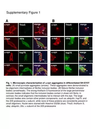
This study presents a detailed microscopic analysis of α-synuclein (α-syn) aggregates in differentiated SH-SY5Y cells. Two distinct types of aggregates were identified: small punctate oligomeric intermediates and mature fibrillar inclusion bodies. The small aggregates do not interact with thioflavin S, while the large inclusion bodies exhibit strong thioflavin S fluorescence, indicating the presence of β-sheet-rich fibrils. Moreover, these large inclusions are associated with proteins such as ubiquitin, Hsp70, and the 20S proteasome α-subunit, which are not found in the small oligomeric intermediates. Nuclei were stained with Hoechst 33258.

Microscopic Characterization of α-Synuclein Aggregates in Differentiated SH-SY5Y Cells
E N D
Presentation Transcript
Supplementary Figure 1 A B merge merge a-syn a-syn ThioS ThioS a-syn ubiq merge ubiq merge a-syn merge a-syn a-syn merge hsp70 hsp70 a-syn a-syn merge merge 20s 20s Fig. 1.Microscopic characterization of a-syn aggregates in differentiated SH-SY5Y cells. (A) small punctate aggregates (arrows). These aggregates were demonstrated to be oligomeric intermediates of fibrillar inclusion bodies. (B) Mature fibrillar inclusion bodies (arrowheads). The strong thioflavin S fluorescence of the large pericentriolar inclusion bodies indicates that the inclusion bodies contain b-sheet-rich fibrils; in contrast, the small oligomeric intermediates fail to interact with this dye. The large inclusion bodies also contain other protein components, such as ubiquitin, hsp70, and the 20S proteasome a-subunit, while none of these proteins are consistently present in small oligomers. Nuclei were stained with Hoechst 33258 (blue). ThioS: thioflavin S, ubiq: ubiquitin, 20s: a-subunit of the 20S proteasome