VLDL formation
VLDL formation. Apolipoprotien B-100 has a repeating - helix/ - sheet structure:. Lipids are packaged as apolipoprotein B-100 is being synthesized:. From Shelness & Sellers (2001) Curr Opin Lipidology 12:151-157. VLDL formation. VLDL stands for Very Low Density Lipoprotein

VLDL formation
E N D
Presentation Transcript
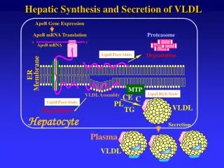
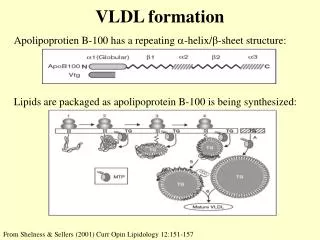
VLDL formation Apolipoprotien B-100 has a repeating -helix/-sheet structure: Lipids are packaged as apolipoprotein B-100 is being synthesized: From Shelness & Sellers (2001) Curr Opin Lipidology 12:151-157
VLDL formation • VLDL stands for Very Low Density Lipoprotein • As it is synthesized, VLDL contains: • One molecule of apoliprotein B-100 • Triacylglycerol • Phospholipid • Cholesterol ester • Microsomal Triacylglycerol Transfer Protein(MTP) assists in the formation of the VLDL • Other components are added to the VLDL in the blood.
VLDL formation • Apolipoprotein B-100 synthesis is required for the transport of lipid out of the liver • If protein synthesis is reduced (e.g. by malnutrition) fat droplets accumulate in the liver. • If the rate of lipid synthesis is greatly elevated with respect to protein synthesis (e.g. in type I diabetes or glucose 6-phosphatase deficiency) fat droplets accumulate in the liver.
Triacylglycerol Oxidation Lipases Triacylglycerol 3 fatty acids + glycerol • During starvation adipose tissue does not release triacylglycerol. • It releases fatty acids and glycerol (produced by adipose lipases). • In the fed state triacylglycerol is transported in the blood • as a lipoprotein complex. In the blood the triacylglycerol • is hydrolyzed to produce fatty acids and glycerol (lipoprotein • lipase or hepatic lipase).
Triacylglycerol Oxidation • Glycerol can be converted to glucose • Glycerol kinase is present in liver but not normally present in adipose Glycerol Glycerol-3-Phosphate DHAP ATP ADP + Pi NAD+ NADH + H + H2C-OH | O=C O | | H2C-O-P-O - || O - H2C-OH | HOCH O | | H2C-O-P-O - || O - H2C-OH | HOCH | H2C-OH Glycerol-3-phosphate dehydrogenase Glycerolkinase
Triacylglycerol Oxidation • Fatty acids must be activated to Acyl-CoA Fatty acid + CoA + ATP Acyl-CoA + AMP + PPi PPi + H2O 2 Pi Acyl-CoA synthetase Pyrophosphatase
Triacylglycerol OxidationRegulation • Fatty acid oxidation takes place in the mitochondria. • Transport into the mitochondria is the primary rate limiting step of fatty acid oxidation. • The maximum rate of fatty acid oxidation is transcriptionally regulated by PPARα. • Unsaturated fatty acids increase PPARα activity • Fibrates, a class of triacylglycerol lowering drugs, increase PPARα activity. • Note PPAR will be persented in Thursday’s lecture
Triacylglycerol Oxidation Inhibited by Malonyl-CoA • Carnitine Shuttle Acyl-CoA + Carnitine Acyl-Carnitine + CoA CAT-I CAT-II CAT-II Acyl-CoA + Carnitine Acyl-Carnitine + CoA Mitochondrion Inner membrane Outer membrane CAT - Carnitine Acyl-CoA Transferase
Triacylglycerol Oxidation • β-oxidation of acyl-CoA • Two carbons at a time are oxidized and removed as acetyl-CoA • For each two carbons removed, 1 FADH2 and 1 NADH + H+ are produced • For palmitoyl-CoA, the reaction is: Palmitoyl-CoA + 7FAD + 7NAD + 7CoA + 7H2O 8Acetyl-CoA + 7FADH2 + 7NADH + 7 H +
Triacylglycerol Oxidation • The first step of the oxidation is catalyzed by Acyl-CoA dehydrogenase. • There are three types, differing in chain length specificity • LCAD - Long chain • MCAD - Medium chain • SCAD - Short chain • In New York State, all newborns are screened for MCAD deficiency • This disorder is covered in the “Baby Ian” case study on the MGB web site.
Triacylglycerol Oxidation • Oxidation of unsaturated fatty acids occurs by the beta oxidation pathway, but with two additional enzymes that isomerize (from cis to trans) and reduce the double bond(s). • Very long chain fatty acids chain fatty acids gre oxidized in peroxisomes to long chain amd medium chain acyl-CoA which enter the mitochondria via the carnitine shuttle. Adrenoleukodystrophy (ALD) is an X-lined disorder in which the entry of very long chain fatty acids into the peroxisome is blocked.
Ketone Bodies • Ketone body synthesis - LIVER (mitochondria) OH O | || CH3-C-CH2-C-S-CoA | CH2 | COO - O || CH3-C-S-CoA CoA O || CH3-C-S-CoA O O || || CH3-C-CH2-C-S-CoA Acetyl-CoA CoA 2 Acetyl-CoA Acetoacetyl-CoA HMG-CoA mitochondrial! Acetyl-CoA NAD+ NADH+H+ OH O | || CH3-C-CH2-CO - O O || || CH3-C-CH2-CO - -hydroxybutyrate Acetoacetate
Ketone Bodies • Acetyl-CoA can be converted into ketone bodies: • Acetoacetate: • -hydroxybutyrate: • These are exported by the liver and used as fuel by other tissues • In a non-enzymatic side reaction, small amounts of acetone are produced from acetoacetate O O || || CH3-C-CH2-CO - OH O | || CH3-C-CH2-CO - O || CH3-C-CH3
Ketone Body Use NAD+ NADH+H+ OH O | || CH3-C-CH2-CO - O O || || CH3-C-CH2-CO - -hydroxybutyrate Acetoacetate NOT present in liver Succinyl-CoA CoA transferase (thiphorase) O || CH3-C-S-CoA Succinate 2 Acetyl-CoA Acetoacetyl-CoA thiolase CoA