Download
1 / 38
420 likes | 595 Vues
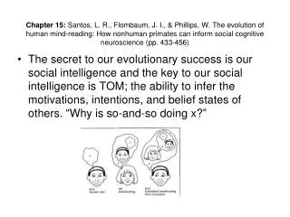
Overview of Neuroscience. Tony Bell Helen Wills Neuroscience Institute University of California at Berkeley. Hubel & Wiesel non-oriented receptive field. Hubel & Wiesel oriented receptive field. Moving epileptic seizure recorded by EEG.

E N D
Overview of Neuroscience Tony Bell Helen Wills Neuroscience Institute University of California at Berkeley
Communication through neuronal coherence (Pascal Fries)
Correlation coefficient between high gamma amplitude and theta amplitude as a function of theta phase. Includes 95% confidence intervals. (Canolty et al, submitted)
Canolty writes: If 1) local pyramidal cell spiking is modulated by the power and phase of local high gamma (neocortical ripples at 80-150 Hz), which are coherent over an area of radius ~0.3-3 mm [14,15], and 2) the power of local high gamma is modulated by the power and phase of the slower theta rhythm (4-8 Hz),which shows a moderate degree of mutual information over a radius of ~ 10-20 mm, then 3) the joint probability density distribution of the conductances of billions of neocortical cells would exhibit a statistical dependence which, without massively parallel recordings, could easily pass for noise -- but which would nonetheless influence computation and cybernetic regulation. This study presents evidence which supports premise 2 of the above argument.
Spikes are not precisely time-locked to the visual stimulus but rather phase-locked to ongoing network activity:
The influence of ongoing oscillations on the spike timing in LGN can be seen in Figure 1. Figure 1a shows the interval distribution for LGN spikes (red)and retinal synaptic inputs (blue) with distinct peaks at certain intervals corresponding to one (or several) periods of the oscillation. Figure 2b shows the cross-correlogram between synaptic inputs (EPSPs) and spikes in LGN. The narrow peak near time 0 shows that LGN spikes follow synaptic inputs with millisecond precision and the side peaks again show periodicity. The inset shows that the oscillations are coherent over several hundred milliseconds. Figure 2c shows that Spikes occur at a preferred phase of the oscillation (upper panel) that has been detected in the synaptic input. The lower panel shows the so-called shift-predictor which quantifies the phase locking of spikes with respect to the previous repetition of the stimulus. The uniform distribution shows that the oscillations are not stimulus locked. In other words, the absolute phase of the oscillations is not determined by the stimulus but rather dynamically generated in the retina. Figure 1c quantifies the amount of phase locking with respect to different frequencies. The phase locking is maximal at frequencies in the gamma range around 60Hz. Another visualization of the influence of ongoing oscillations on spike timing in LGN: Figure 2a shows spike trains (red) and synaptic input (EPSP) trains (blue) in response to the onset of the movie stimulus at 2.0s. The curve in in the upper panel is the average response or PSTH (post stimulus time histogram). It can be seen that even though the response rate is reproducible from trial to trial, the spike timing on the millisecond scale is seemingly random. Figure 2b shows the same activity after each trial has been re-aligned by a time shift corresponding to the oscillation phase that we could determine from ongoing activity in the input preceding the stimulus (blue). Now the spike in response to the stimulus (red) line up on a millisecond scale. Thus, the spikes are not precisely time-locked to the visual stimulus but rather phase-locked to ongoing network activity!
Conclusion. Neurobiology consists of nested networks of diverse mechanisms. There is no “magic level” like the spiking level or the rate level. Information can flow very quickly up and down through the levels. Up through emergence. Down through boundary condition. These cross-level connections form and dissolve dynamically. This is regulated by ‘coherences’ at a given level. Thus it is a ‘dance’ of intra-level and inter-level resonances and information flows. Question. Is there an invariance to how this dance self-organises? Can we find that invariance and create a theory of complex systems? To stand a chance of answering this question, we will need to do some mathematics. Starting tomorrow…