Palliative Wound Care
Palliative Wound Care. Helping when it won’t heal September 28, 2011 Kathleen Kelly MD, CWS-P katkelly@ehr.org. I: Basic principles for wound care. Control and eliminate causative factors Provide systemic support to eliminate potential and existing cofactors

Palliative Wound Care
E N D
Presentation Transcript
Palliative Wound Care Helping when it won’t heal September 28, 2011 Kathleen Kelly MD, CWS-P katkelly@ehr.org
I: Basic principles for wound care • Control and eliminate causative factors • Provide systemic support to eliminate potential and existing cofactors • Maintain a physiologic local wound environment
Manage: pain Odor Bleeding exudate Maintain QOL for patient and caregiver Palliative wound care goals
Bottom line • Clean it • Cover it • Moisten it • Waterproof surrounding tissue • Protect it • Feed it
Skin • Protection from external elements • Fluid and electrolyte homeostasis • Immunological response • Waterproofing with sebum • Melanin for solar protection • Environmental sensation • Thermoregulation • Emotions and body image • Vitamin D metabolism
Epidermis—mostly dead Waterproofing Sun protection Cohesion Dermis Nerves Sweat/oil glands Hair Subcutaneous Loose Fat Lymphatics blood vessels Fascia Muscles Bone Skin Structure
Cascade of normal healing • Hemostasis stop bleeding • Inflammation clean out • Proliferation fill in • Maturation reorganize for strength • Max tensile strength 80%, takes 2 yrs
ACUTE 24 hrs to mobilize epis 7 days to new vessels & fibroblasts 9 days to collagen RE-epithelialize CHRONIC Impaired inflam phase Meds Nutrition Immune status Stuck in inflam phase Acute vs Chronic Wounds
Obstacles to Healing“Mr. Fetid” • Maceration, malnutrition • Radiation • Foreign body (drainage, necrosis), Fistula • Epibole (rolled edge)/epithelial invasion • Tumor, trauma, toxin (H2O2!) • Infection, ischemia • Drugs (steroids, CTX)
Age Anticoagulation PVD Anemia Renal insufficiency AIDS Infection DIABETES Malnutrition Transplant status Chemotherapy Steroids NSAID States a/w delayed healing
The scary past 1650 BC Egyptians use the first bandages • Linen strips • Lint with medicine for topical care 3400 yrs of dry dusty dressings 1962 Plastic films and non-woven dressings healed wounds twice as fast as allowing scab to form (pigskin model)
Water Vapor Loss • Across intact skin: 42 gm per day per sq m • ~1/3 cup • Across denuded skin: 7,874 gm per sq m • 2 gallons
OPEN wound • Dehydration necrosis of superficial cells forms scab –bacterial hideout • Epis migrate only over viable (moist) tissue • Evaporative cooling loses 4 hrs healing time per dressing change • Cool tissue increases risk for infection. (vasoconstriction = less tissue O2)
Ideal conditions for cell activity • Normal temp • Moist environment for O2 and nutrients • Minimum disturbance • Exudate/edema control • Adequate host nutrition
Smoking • Nicotine causes vasoconstriction for up to 50 minutes • CO forms carboxyhemoglobin, makes platelets stick • Increases blood viscosity, aggravates ischemia • Depletes vitamin C
II: How to Evaluate • Standard language for reporting what you find • Rationale for deciding what to do about it
Wound measurement • Anatomic position • Clean wound with saline first • Length from 12:00 to 6:00 • Width from 3:00 to 9:00 • Depth from visible surface to max depth
Granulation: looks like raw burger Hypergranulation: sticking up Slough: nonviable biomatter Eschar (scab) Epithelial Muscle/tendon/fascia/bone Adherence Color Odor Foreign bodies Exudate Wound Bed Tissue Type
THE GOOD Flush Carrier medium for repair supplies & cells Lube for epi migration THE BAD Excess Maceration Proteolysis Insufficiency Stuck bandage accumulated slough Exudate (coffee for skin)
Edges • Defined/undefined • Attached/unattached • Fibrotic/callused • Macerated/soft • Rolled (epibole) • Tunneling/undermining
Erythema Edema Crepitus Color Texture Maceration Temperature Scar Xerosis Ecchymosis Rash Surrounding Tissue
Wound assessmentThe system (whole), not the hole • Duration (acute vs. chronic) • Cause • Environmental factors –tx to date • Systemic factors –can the pt heal? • Psychosocial • PAIN
Bacteria in woundspenetrate 64 layers of gauze • Contaminated: All wounds! • Not multiplying=no effect on healing • Colonized • Replicating but no injury to host • Critically colonized • Replication starting to injure local tissue • Infected • Invasion to healthy tissue
“Classic” Induration Fever Edema erythema “New” Tissue breakdown Pain Exudate Discoloration Delayed healing Odor Infection signs
Topical antiseptics-cytotoxic • Burow’s solution (aluminum salt) • Betadine, crystal violet • MRSA, fungi, virus, protozoa • Dakin’s • 1/4s--burns • Acetic acid • Pseudomonas—apply TID • H2O2 • Cytotoxic 1000 x past the end of bactericidal activity
Topical antibacterialsprevent multiplication • Iodoflex (cadexomer iodine) • Change 3/7; goes on brown, turns yellow-gray • Silver • Compounded with multiple dressings • Hydrofera Blue • Gentian violet – change when it turns white
Mupirocin –MRSA Polymyxin—gram neg Bacitracin –gram pos CONTACT DERMATITIS Limit to 2 weeks Don’t use same po and topical Topical antibioticskill bacteria
III: Wound bed preparation: TIME • Tissue management (get rid of garbage) • Debridement, foreign matter • Inflammation and infection control • Moisture Balance • Epithelial edge advancement
Dressing goalsExudate, wound bed, pain • Remove excess exudate & toxins • Maintain high humidity @wound/dressing interface • Allow gas exchange • Thermal insulation • Protect from secondary infection • Prevent contamination • Allow trauma free removal at dsg change
Design your dressing • Contact layer: so it doesn’t hurt to change the dressing • Space filler: for deep wounds/tunnels • Moisture balancing layer • Protect surrounding skin • Something to hold it on • Someone to change it/bathing rules • Pain management please • Address underlying cause
Heavy drainage managementChange for strike-through Kurt’s sock • Alginate (seaweed) • Hydrofiber (felt to slug) • Foam (sponge) • Pouch (stoma bag) • Protect surrounding skin (paste)
Topical formulations • Lotion: powder in water • Cream: oil in water • Ointment: water in oil • Paste: powder in ointment • Gel: particles in lattice
Moisture donationfor dry beds: daily change • Amorphous gel –solosite or duoderm gel (goo) • Vaseline gauze/xeroform/adaptic • Hydrogel sheet –curagel (jello) • Hydrocolloid sheet –duoderm (cheese) • Occlusive dressing – tegaderm (saran)
Dead space • Chicken soup • Filling promotes healing from base of wound • Avoids abscess formation • ONE piece of filler, & anchor it • Choose nonstick contact layer based on bed moisture • Protein nutrition
Stove-top Saline • 2 tsp salt in 1 liter H20 • Boil 3-20 min • Irrigate < 15 PSI • 20g angiocath + 30-60 cc syringe
tape • Remove toward wound • Apply parallel to incision • Clean dry skin (clipped, not shaven) • Skin prep • Don’t encircle limb • Use stretchy tape if distension anticipated. • Extend at least ½ inch beyond dressing • Fold end over for easy removal
Mesalt hypertonic saline Iodoform iodine nugauze Xeroform Vaseline/bismuth Vaseline occlusive Adaptic Oil emulsion Kerlix AMD Antimicrobial Kerlix fluffy Kling Stretch net Gauze
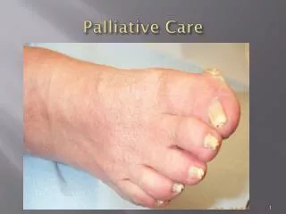
The wounds we see most • Venous • Arterial • Diabetic/neuropathic • Pressure ulcers • Skin cancer
More common palliative wounds • Pressure ulcers • Arterial • Neoplastic • Venous insufficiency • Kennedy
Pressure ulcers • Stage I: NOT Blanchable, red, squishy, intact • Stage II: Partial thickness:pink, partial, painful blister or crater • Stage III: Full thickness as deep as fascia • Stage IV: deeper than fascia • Unstageable: base covered by eschar/slough • NO REVERSE STAGING • Deep Tissue Injury • Kennedy ulcer
Kennedy Ulcer • Pressure ulcer of dying • Looks like dirty skin • Blister (st II) with rapid development to St III-IV • Not preventable • Treat as other PU: odor and drainage control, no debridement
Arterial wounds • Get ABI: Artifactually high in DM pts with calcified vessels -- Try TBPI or Toe pressure. Normal >=1; Severe ischemia <= 0.5 • Dry stable eschar – LEAVE IT!!!! • If the scab oozes, lose it. • Daily dressing
Venous insufficiencyConfirm artery supply 1st • Compress if ABI >=0.8 • Unnas work only in ambulatory patients • Teds are for beds. • Multi-layer wraps • tubigrips • Intermittent compression pumps • Tubi-grips
Oddballs • Kennedy ulcer • Kaposi Sarcoma • Pyoderma Gangrenosum –pathergy • Fungating • Marjolin squamous ca at scar site • Necrotizing Fasciitis --DM
Attachments • Dressing instruction checklist • Whadaya call it • Hospital order sheets online • Stage I & II • Stage III & IV, Venous, arterial, neuropathic • VAC