
Stem Cells
E N D
Presentation Transcript
The Fertilized Egg is The Ultimate Stem Cell Comparison of Normal Development, Reproductive Cloning & Therapeutic Cloning Hochedlinger, K. & Jaenisch, R. N Engl J Med 2003; 349:275-286
Dolly! Dolly at her press conference explaining her importance
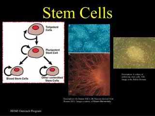
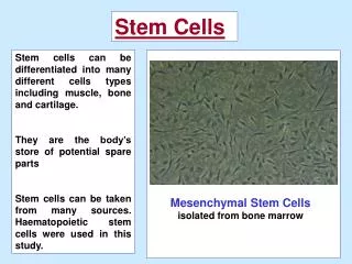
Many different Cell Types Arise From 3 Germ Layers Epithelia
Early Development Generates a Pluripotent Stem Cell Population The Generation of Embryonic Stem Cells after Somatic-Cell Nuclear Transfer Red & White blood cells Snyder, E. Y. et al. N Engl J Med 2006;354:321-324
Cardiomyocytes From Human ESCs The scheme shows the directed differentiation of human ESCs to cardiomyocytes and their application for cardiac repair in a rat model of cardiac infarct (Laflamme et al., 2007). Undifferentiated human ESC colonies are replated as high-density monolayers, expanded, and then induced to differentiate by sequential treatment with activin A (day 0) and BMP4 (day 1). Differentiation along the cardiac lineage can be further enhanced by activating the Wnt/β-catenin pathway. Cultures typically exhibit vigorous beating activity 10–14 days postinduction. These populations are then subjected to heat shock and treated with IGF-1 24 hr prior to transplantation to enhance viability, and then enriched for cardiomyocytes using Percoll density-gradient centrifugation. They are then suspended in a “prosurvival cocktail” to block cell-death pathways, and are delivered to the infarcted heart by direct injection. Experimental endpoints are assessed by microscopy and magnetic resonance imaging. By combining these advances, researchers have generated significant amounts of human myocardium in the infarcted rat heart, reaching up to 11% of the infarct's volume (Figure 4) (Laflamme et al., 2007). The human myocardium prevented the progression to heart failure seen in untreated rats and in control animals receiving noncardiac derivatives of human ESCs.
(A) Shown is a confocal fluorescent micrograph of a human myocardial graft in an infarcted rat heart. The peri-infarct zone is stained with human-specific β-myosin heavy chain (red) and pan-cardiac marker cardiac troponin I (green) revealing immature human cardiomyocytes (yellow) in close apposition to host cardiomyocytes (green). (Nature Biotechnology 25, 1015, 2007). (B) Human cardiomyocyte engraftment and cardiac contractile function. Magnetic resonance imaging demonstrates a 2.5-fold enhancement of systolic wall thickening in the infarct region of the rat heart receiving a human cardiomyocyte graft. Control groups received noncardiac human ESC derivatives in prosurvival cocktail (PSC), PSC only, or serum-free media (SFM only). NS, no significant difference. (Adapted from Laflamme et al., 2007 M.A. Laflamme et al., Biotechnol. 25 (2007)
Strategies to Induce Reprogramming of Somatic Cells(1) Nuclear transfer involves the injection of a somatic nucleus into an enucleated oocyte, which, upon transfer into a surrogate mother, can give rise to a clone (“reproductive cloning”), or, upon explanation in culture, can give rise to genetically matched embryonic stem (ES) cells (“somatic cell nuclear transfer,” SCNT). (2) Cell fusion of somatic cells with ES cells results in the generation of hybrids that show all features of pluripotent ES cells. (3) Explantation of somatic cells in culture selects for immortal cell lines that may be pluripotent or multipotent. At present, spermatogonial stem cells are the only source of pluripotent cells that can be derived from postnatal animals.
Yamanaka’s Bombshell Takahashi & Yamanaka Cell 2006
Characterization of iPS Cells Derived from Adult Mouse Tail-Tip Fibroblasts(A) Morphology of iPS-TTFgfp4-3 on STO feeder cells.(B) RT-PCR analysis of ES marker gene expression in iPS-TTFgfp4 cells (clones 1–5 and 7). We used primer sets that amplified endogenous but not transgenic transcripts.(C) Contribution of iPS-TTFgfp4-7 and iPS-TTFgfp4-3 cells to mouse embryonic development. iPS cells were microinjected into C57/BL6 blastocysts. Embryos were analyzed with a fluorescence microscope at E7.5 (upper panels, iPS-TTFgfp4-7) or E13.5 (lower panels, iPS-TTFgfp4-3). Scale bars = 200 μm (upper panels) and 2 mm (lower panels).(D) The E13.5 chimeric embryo was sectioned and stained with anti-GFP antibody (brown). Cells were counterstained with eosin (blue).
But Expression of Some Genes May be Better Indicators than Others Mice from iPS cells
Derivation of autologous iPS cells from hβS/hβS mice and correction of the sickle allele by gene targeting. (A) Scheme for in vitro reprogramming of skin fibroblasts with defined transcription factors combined with gene and cell therapy to correct sickle cell anemia in mice. (B) Representative images of various steps of deriving hβS/hβS iPS line #3. (C) Southern blot for c-Myc viral integrations in (i) ES cells, (ii) hβS/hβS iPS line #3 and (iii) its derived subclone hβS/hβS iPS #3.3 obtained after infection with adeno-Cre virus and deletion of the viral c-Myc copies. * indicates endogenous c-Myc band. Arrows point to transgenic copies of c-Myc. (D) hβS/hβS iPS#3.3 displayed normal karyotype 40XY (upper left), was able to generate viable chimeras (upper right), and formed teratomas (bottom). (E) Replacement of the hβS gene with a hβA globin gene in sickle iPS cell line #3.3. Homologous recombinants were identified by PCR to identify correct 5' and 3' end replacement. PCR with primers 3 and 4 followed by Bsu36I digestion was used to distinguish hβS and hβA alleles. Correctly targeted clone #11 displayed identical pattern to that previously obtained for correctly targeted ES cell clone
a, Schematic diagram of the experimental strategy. Adenoviruses encoding bicistronic transcription factor (TF) and nGFP linked by an IRES element (I) were injected into the pancreas of an adult mouse (Rag-/-). CMV, cytomegaloviral promoter. b, Wild type (WT) pancreas is predominantly exocrine tissue with insulin+ -cells in the islet (outlined). Nuclei were stained blue with DAPI. c, One month after infection with a combination of Ngn3, Pdx1 and Mafa viruses (pAd-M3), numerous insulin+ cells appear outside of islets. d, e, Quantification of induction one month after infection. M9, M6: mixture of 9 and 6 different viruses, respectively. Data are presented as mean + s.d.; n = 3 animals. 1,000 nGFP+ cells were counted per animal. Asterisk, P < 0.05; two asterisks, P < 0.01; three asterisks, P < 0.001.