INCONTINENCE
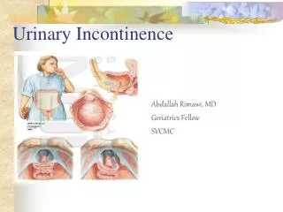
INCONTINENCE. Defined as “the involuntary loss of urine due to increased bladder pressures or decreased ability of the vesicourethral sphincter to prevent the escape of urine” (Porth 2002). http://www.continence-foundation.org.uk/publications/.

INCONTINENCE
E N D
Presentation Transcript
Defined as “the involuntary loss of urine due to increased bladder pressures or decreased ability of the vesicourethral sphincter to prevent the escape of urine” (Porth 2002).
http://www.continence-foundation.org.uk/publications/ Despite popular opinion, incontinence as part of the normal aging process is not the only cause. • 10-25% of women between the ages of 15 and 64 experience incontinence at some time. • It affects over 30% of community - living adults • 60% of residents in long-term aged care facilities.
It affects: • Teenage athletes • Pregnant women – 50% of women who have had children develop a prolapse. • New mothers • Menopausal women 45–60 years old • Women after gynaecological surgery • Only 10–20% actually seek help • It has been estimated that urinary incontinence costs the USA around $15 billion per year.
Causes INHERITED OR GENETIC FACTORS • Anatomic difficulties • Weakness of muscles that hold the bladder in place or in the bladder itself • Weakness of the urethral sphincter muscles • Overactive bladder muscles. • In men, a non-cancerous overgrowth of the prostate gland, prostate cancer or prostate surgery.
Neurological abnormalities • Damage to the nerves controlling the bladder as a result of diseases such as diabetes, stroke, Parkinson's disease and multiple sclerosis. • Hysterectomies and prostatectomies can weaken or damage the muscles and nerves of the urinary tract and can cause incontinence. PHYSICAL FACTORS • Pregnancy and childbirth • Pressure of the uterus and contents • Connective tissue changes or injury to the fascia • Mechanical disruption of muscles and sphincters
Aging • Hormonal effects • High concentration of oestrogen receptors in pelvic tissue. • General collagen deficiency state or a decrease in estrogen in post-menopausal women can reduce the strength of the sphincter muscle. • Urethral coaptation affected by the loss of oestrogen • Non-obstetric pelvic trauma and / or surgery • Medications
TYPES STRESS INCONTINENCE • Involuntary loss of urine during coughing, sneezing, laughing or other physical activities that increase intra-abdominal pressure. • Caused by • a weakness of the pelvic floor muscles • Structural damage to the bladder neck or urethral sphincters following surgery or trauma • Breakdown in bladder support secondary to birth, trauma or age • Most common in younger premenopausal women
In men can be caused by • congenital defect • Trauma • Surgery to the bladder outlet such as prostatectomy URGE INCONTINENCE (Also called detrusor instability or an overactive bladder) • Hyperactivity of the bladder characterised by spontaneous, uninhibited bladder contractions with an urgency to void. • May or may not involve involuntary loss of urine
Usually idiopathic although it can be linked to • underlying neurological disease such as • Stroke • Parkinson’s disease • Multiple sclerosis • Injury • Aging • Radiation to the lower abdomen • Chronic bladder outlet obstruction causing a partial destruction of the nerve endings that control bladder excitability • Inflammation or irritation of the bladder
Disease conditions such as diabetes mellitus • Drugs such as hypnotics, tranquillisers and sedatives can interfere with the conscious inhibition of voiding, leading to urge incontinence • Diuretics also increase the flow of urine and may contribute to urge incontinence • More common in older post-menopausal women
OVERFLOW INCONTINENCE • Loss of urine involuntarily secondary to a failure to empty • Bladder capacity is reached then the bladder pressure becomes so great that it overcomes any resistance to the outlet • Symptoms • Frequent or constant dribbling • Urge / stress incontinence symptoms • Recurrent urinary tract infections
Causes • Decreased bladder contractility • Age • Bladder over-distention • Severe infections • fibrosis • Bladder outlet obstruction • Large cystoceles • Urethral strictures • Closed sphincters
Medications • Diuretics causing the bladder to fill more rapidly than usual • Anticholinergics inhibit overactive bladder contractions • Psychotropics • Alpha-adrenergic blockers relax smooth muscle in the bladder neck and prostatic urethra, decreasing urethral resistance
In males, one of the most common causes of this is enlargement of the prostate gland • Another cause often overlooked is faecal impaction. When a large bolus of stool forms in the rectum, it can push against the urethra and block the flow of urine
FUNCTIONAL INCONTINENCE • Results from a deficit in the ability to perform toileting secondary to factors outside the urinary tract e.g. chronic physical or mental impairment REFLEX INCONTINENCE • Unconscious incontinence • Common in paraplegics
TREATMENT PROTOCOL For stress incontinence associated with mild vaginal prolapse, herbs which boost oestrogenic activity for the premenopausal woman as well as post menopausal indications • Discorea villosa • Chamaelirium lutuem • Tribulus terrestis • Is indicated for many conditions from impotence to urolithiasis and incontinence. • Lignans (linseed)
Tone the bladder sphincter to reverse bladder leakage problems • Equisetum arvense • Hemostatic and collagen astringent for connective tissue weakness accompanying nephritis, mucosa astringent in UTI. (Moore, M. “HERBAL TINCTURES IN CLINICAL PRACTICE, 2.0” Southwest School of Botanical Medicine) • Agropyron repens • Serenoa repens • Crataeva nurvala • Provides relief from incontinence, frequency and pain and an improvement in bladder tone. (Bone. K. 1997. “Clinical Application of Ayurvedic and Chinese Herbs”, Phytotherapy Press, Warwick) • Agathosma betulina
Soothe and heal the urinary tract • Barosma betulina • Inflammation of the urinary tract • Cystitis, urethritis, dysuria with urging but little relief (Moore, M. “HERBAL TINCTURES IN CLINICAL PRACTICE, 2.0” Southwest School of Botanical Medicine) • Galium aparine • Agropyrens repens • Any acute irritation of the urinary tract or kidneys, with referred pain along flanks, pain on urination; soothing, not disinfectant. (Moore, M. “HERBAL TINCTURES IN CLINICAL PRACTICE, 2.0” Southwest School of Botanical Medicine) • Arctostaphylus uva-ursi • Inflammation of the urinary tract and bladder (Wichtl & Bisset 2000)
Zea Mays • Equisetum arvense • A diuretic that increases the flow through the ureters, and is useful in inflammation (Wichtl & Bisset 2000) • Althea officinalis radix Boosting the circulation to the pelvic region • Dong Quai • Ginkgo
Crystalline structures that form from components in the urine. • Aetiology of urinary stone formation is complex and is thought to involve a number of factors such as • Increases in blood and urinary levels of stone components • Anatomic changes in urinary tract structures • Metabolic and endocrine influences • Dietary and intestinal absorption factors • Urinary tract infections • Low carbohydrate diets present a possible risk of kidney stones and kidney disease
Kidney stones have an estimated Australian prevalence rate of (Kron, J. 2008, ‘Kidney Stones’, The Journal of Complementary Medicine, vol.7, no.6, pp.14-16) • 6 – 9% in men • 3 – 4% in women • Recurrence rate of 26 – 53% after 10 years
Three theories exist • The Saturation Theory the risk of stone formation is increased when urine is supersaturated with stone components. The greater the concentration, the more likely to find precipitation • The matrix theory organic materials, such as polysaccharides can be found in all layers of kidney stones and either contribute to the initiation of the stone formation or the material is entrapped as the stone forms • The inhibitor theory people who have a deficiency of proteins that inhibit stone formation in their urine are at increased risk for stone formation. Much of the information about organic inhibitors in terms of stone formation is still experimental.
Types of stones: Calcium stones (70-80%) • Usually occur as calcium oxalate and less commonly as calcium phosphate • Associated with increased concentrations of calcium in the blood and urine • Excessive bone resorption can be associated with • Immobility • Bone disease • Hyperparathyroidism • Renal tubular disorders resulting in excessive leakage of clacium • High oxalate concentrations in the blood and urine
Calcium containing calculi have been shown to develop in up to 6% of people treated with indinavir, a protease inhibitor used in HIV infection • About 50% of patients with calcium stones have excessive calcium in their urine. The most common cause of this is a genetically-determined increased calcium absorption in the intestine.
Other contributing factors could include • Excessive urinary calcium can be caused by a diet rich in sodium or animal protein or low in fluid intake. • Parathyroid hormone and vitamin D excess can lead to hypercalcaemia and kidney stones. • Low levels of citrate (affecting between 20 and 60% of patients) • Chronic diarrhoea • Potassium loss • Excessive physical exercise • A high acid forming diet (rich in high protein foods)
Magnesium ammonium phosphate stones (Struvite stones) • Form in alkaline urine and in the presence of bacteria that possess an enzyme called urease, which splits the urea in the urine into ammonia and carbon dioxide. • The increased ammonia helps to increase the pH of the urine, making it more alkaline. • These stones enlarge as the bacterial count grows and they can increase in size until they fill the entire renal pelvis. • Because of their shape, they are often called staghorn stones. • Treatment of the infection is often difficult. • Struvite stones are usually too large to pass and require surgical removal or lithotripsy.
Uric acid stones • Develop in conditions of gout and high concentrations of uric acid in the urine. • High uric acid levels contribute to calcium stone formation by acting as a nucleus for calcium stone formation • They form in urine with a pH of 5.1 to 5.9 • A contributing factor could be a high purine diet Cystine stones • Lithotripsy Shock wave lithotripsy (SWL) uses high frequency sound waves from an external source (outside the body) to break a kidney stone into small pieces, and allow it to pass through the urinary tract.
Manifestations Pain • Renal colic • Caused by stones 1–5mm in diameter that move into the ureter and obstruct flow • Acute, intermittent and excruciating pain in the upper quadrant of the abdomen on the affected side • The pain may radiate to the lower abdominal quadrant , bladder area, perineum or scrotum in the male • The skin may be cool and clammy • Nausea and vomiting are common
Non-colicky renal pain • Caused by stones that cause distension of the renal calculi or renal pelvis • The pain is usually a dull, deep ache in the back that can vary in intensity from mild to severe • The pain may be exaggerated by drinking large amounts of fluid Blood in the urine Increased frequency of urination Pain and burning during urination Fever, chills, loss of appetite Urinary tract infection
Treatment protocol Prevention of kidney stones and the treatment of existing stones • Crataeva nurvala • A clinical study on the effects of Crataeva on 46 patients with kidney, ureter or bladder stones not requiring surgery found that 26 patients passed the stones within 10 weeks of treatment. The majority of the remaining patients experienced symptom relief. (Bone. K. 1997. “Clinical Application of Ayurvedic and Chinese Herbs”, Phytotherapy Press, Warwick) • Shows a decreased tendency to form calcium oxalate kidney stones. In an in vivo study, it was found that the weight of stones decreased, the bladders of rats showed less ulceration and less cellular infiltration when compared to controls. (Bone. K. 1997. “Clinical Application of Ayurvedic and Chinese Herbs”, Phytotherapy Press, Warwick)
Equisetum arvense • One of the richest sources of natural silica, a mineral which helps healthy calcium metabolism • Supports healthy urine production • German Commission E lists it for static oedema, bacterial infections and inflammation of the lower urinary tract and for renal gravel. • Eupatorium purpureum • Traditionally used as a treatment for stones and gravel in the kidneys • Taraxacum officinalis folia • Grieve (1973) states that it is “a strong decoction that is found to be servicable in stone and gravel”
Hydrangea aborescens • Pyelitis, urethritis, prostatitis; to reduce ureter colic pain and mucosa inflammation (Moore, M. “HERBAL TINCTURES IN CLINICAL PRACTICE, 2.0” Southwest School of Botanical Medicine) • Inflammation of the bladder, urethra and prostate and kidney stones. (Bone, K. 2005) • Sedative effect on the urinary tract • Used in the treatment of kidney stone • Capsella bursa • Urinary astringent in urethritis and the prevention of urate stones.
Pain relief • Viburnum opulus • Relaxes smooth muscle • antispasmodic • Dioscorea villosa • Corydalis ambigua • Almost any type of pain, but particularly visceral pain. It strengthens the analgesic function and has a sedative action as well. (Bone. K. 1997. “Clinical Application of Ayurvedic and Chinese Herbs”, Phytotherapy Press, Warwick) • Caulophyllum thalictroides • Used specifically for smooth muscle spasm and cramping (Bone, K. 2003)
Damage to the urinary tract mucosa • Immune enhancing • Demulcents • Zea mays • Althea officinalis radix • Licorice • Anti-inflammatory action – in vitro studies have shown it has mucoprotective effects allowing tissue and gut repair (Braun & Cohen, 2005) • Urinary tract antiseptics • Arctostaphylos uva ursi
Binding of calcium in the urine and making it less likely to precipitate – anthraquinone containing herbs • Cascara • Yellow dock • Infection as it can be a cause of stone formation • Galium aparine • Echinacea spp.
Increasing fluid intake will help to prevent stone formation by reducing crystal salt concentration and increasing their expulsion. (Kron, J. 2008, ‘Kidney Stones’, The Journal of Complementary Medicine, vol.7, no.6, pp.14-16) • The recommendation is to drink enough water to produce 2 – 2.3 litres of urine each day. Other beverages are additional to water. • No RCTs have been done for specific beverages, however, non-controlled studies have found that orange juice and lemonade increase urinary pH levels and citrate excretion. • Prospective studies have found that coffee, beer and wine are associated with reduced stone formation risk due to increased urinary volume, whereas grapefruit juice and apple juice are associated with an increased risk, although the reason why is not know.
(Kron, J. 2008, ‘Kidney Stones’, The Journal of Complementary Medicine, vol.7, no.6, pp.14-16) makes the following recommendations: • Protein Animal protein (meat, poultry and fish) is regarded as increasing stone-formation risk due to increased urinary calcium, uric acid and phosphate excretion and reduced citrate and urine pH. The animal protein recommendation is 20g/day • Salt mixed results from cohort studies but it is felt that salt is associated with increased stone formation due to increased urinary calcium and uric acid excretion and decreased citrate excretion. • Dairy a high calcium intake is regarded as decreasing stone formation risk by reducing gut absorption of oxalates through calcium binding to them.
Increased vitamin D intake combined with calcium supplementation increases calcium excretion due to excessive blood calcitriol or overregulation of its receptors, leading to excessive absorption of gut calcium. • It is recommended that vitamin D supplementation be avoided except in people with hypovitaminosis D • Magnesium is regarded as decreasing gut absorption of oxalate. A systemic review found that the addition of magnesium to potassium citrate therapy improves outcomes.
Lifestyle(Bone, K. 2004) • Regular weight bearing exercise will help store calcium in bones, which would otherwise be excreted in the urine. However, be aware that excessive exercise increases dehydration and cause lactic acidosis, a factor in stone formation. • Avoid commercial drinks as they are often loaded with phosphate and sugar. • Diet should be based on fruit, vegetables and unrefined carbohydrates. Fruits rich in potassium and citrate should be emphasised together with foods rich in magnesium such as fermented soya products, legumes, nuts and green leafy vegetables.
Calcium intake should not be excessive but should also not be restricted. Restriction of calcium can lead to excessive oxalate absorption • If there is a history of oxalate stones, then oxalates are to be avoided rhubarb, spinach, strawberries, ginger, almonds, cashews, beetroot, tea and chocolate
The average pH of soft drinks, e.g. Coke & Pepsi is 3.4. This acidity is strong enough to dissolve teeth and bones! Our human body stops building bones at around the age of 30. After that, it shall dissolve the bones every year through the urine depending on the acidity of the food intake. All the dissolved calcium compounds, get accumulated in the arteries, veins, skin, tissue and organs, which affects the functioning of the kidney assisting in formation of kidney stones.