Understanding Cell Structure and Function: Discovery, Types, and Organelles
This chapter delves into the intricate world of cells, highlighting their foundational role in life. It explores the history of microscopy, beginning with early prototypes and pivotal discoveries by scientists like Robert Hooke and Anton van Leeuwenhoek. The cell theory is outlined, emphasizing that all living organisms are composed of cells. The text discusses various microscope types, including light, dissecting, and electron microscopes, as well as the characteristics of prokaryotic and eukaryotic cells. Additionally, it examines cellular organization, focusing on organelles, the nucleus, and cellular processes.

Understanding Cell Structure and Function: Discovery, Types, and Organelles
E N D
Presentation Transcript
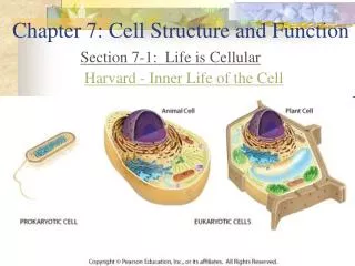
Chapter 7 Cell Structure and Function Chapter 7 Cells 2010-2011
I. 7.1 Life is cellular • The Discovery of the cell • Early microscopes a. Prototypes were developed in the late 1500’s by European eyeglass makers Early compound microscope 17th century
b. In 1665, Robert Hooke used an early compound microscope to look at cork • Observed that cork was made up of thousands of hollow chambers • Dubbed them cells since they looked like the monatsery’s tiny rooms called “cellula”.
c. Late 1600’s- Dutch textile salesman: Anton van Leeuwenhoek • Created different types of microscopes • Discovered over 5,000 types of microscopic life • Lenses were able to magnify up to 300X
2. The Cell Theory- • Cells are the most basic unit and structure of life • All living things are made up of cells • New cells are produced from existing cells
B. Exploring the Cell 1. There are 3 major types of microscopes • a. Light Microscope • Specs. • Magnifies 40 – 1,000 times depending on objective being used • Eyepiece magnifies 10X • Total magnification calculation: multiply the eyepiece by the objective being used. Example- eyepiece (10X) times low power objective (4X) = 40X • Used to magnify objects that light can pass through. • Uses slides
b. Dissecting Microscope Specs. • Magnifies 10 to 30 times • Eyepiece magnifies 10X • Objectives: 1X and 2X (3X) • Used to magnify objects that light cannot pass through • Used mostly by research scientists and jewelers • Advantage: objects are 3-D • Disadvantage: can’t view small objects
c. Electron Microscope • Uses electrons to illuminate objects • Can magnify from 30,000 to 9 million times • Two types: Transmission and Scanning • Mostly large institutions have them • Costly to own and maintain • Can only be used to look at dead specimens • Used for cytology, forensics, and virology
Transmission Electron Microscope • TEM- thin slices need to be made to have clear images, images are 2-D • Useful for studying internal structures
SEM- samples do not need to be cut, are in 3-D • Useful for studying external structure
C. Prokaryotes and Eukaryotes • General Information on cells • Cells come in a variety of shapes • Range in size from microscopic bacteria to giant amoeba Mycoplasma pneumoniae Chaos carolinensis – Giant amoeba, approximately 1mm in length
c. All contain DNA d. All have a cell membrane- an outer, flexible barrier Cell membrane Nucleus- containing DNA
2. Two Main Categories of Cells • Eukaryotes- have cells that enclose their DNA in a nucleus • Prokaryotes- cells that do not enclose their DNA in a nucleus
3. Prokaryotes • Are generally smaller than eukaryotic cells • Have no nucleus • Carry out all of life’s processes • Ex: bacteria Bacillus anthracis
4. Eukaryotes • Are generally larger and more complex than prokaryotes • Contain dozens of membrane bound structures that are specialized • Nucleus separates DNA from rest of cell
d. Come in a variety of shapes and size e. Ex: protists, fungi, plant, and animal cells
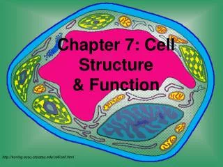
II. 7.2 Cell Structure • Cell organization 1. Organelles- “tiny organs”; the specialized structures within the cell that perform a function
2. Cytoplasm- the portion of the cell outside of the nucleus; the fluid that fills the entire cell • Makes up about 70% of the cells volume. • Most of the cells chemical reactions occur here
3. The cell as a factory • The different organelles of a cell can be compared to a living version of a modern factory • Cells, like factories follow instructions to produce products
4. The Nucleus- the “control center” of the cell • Contains nearly all of the cells DNA and, with it, the coded instructions for making proteins and other important molecules • Prokaryotic cells lack a nucleus, but they do have DNA
c. Parts of the nucleus • Nuclear membrane- aka “nuclear envelope”; composed of two membranes; allows for materials to come in and out through pores • Nuclear pore- allows proteins, RNA, and other molecules to move in and out of the nucleus
Inside the Nucleus • Contains the nucleolus- darkened area in nucleus that directs the formation of ribosomes • Chromosome- carries the cells genetic information; important for cell division
B. Organelles that Store, Clean-up, and Support • Vacuoles- large sac-like, membrane enclosed structures • Function- store water, salts, proteins, carbohydrates • Larger in plant cells
c. Found in some unicellular organisms and animal cells • Ex: Paramecium Contractile vacuole in paramecium cell
d. Typically called vesicles in animal cells- store and move materials between cell organelles and to the cell surface
2. Lysosomes- the “clean-up crew”; small organelles filled with enzymes • Break down many types of materials • Lipids • Carbohydrates • Proteins • Cellular debri b. Found mostly in animal cells
Lysosomes (cont) • Found mostly in white blood cells • Have been linked to diseases, such as Tay Sach’s • Tay Sach’s is a disorder that is caused by a genetic defect that prevents the formation of an essential enzyme that breaks down lipids • These lipids build up in the body and can cause nerve damage; prognosis is not good
3. Cytoskeleton • The framework of the cell ; a network of protein filaments • Some parts help in transport of materials • Aids the cell in movement • Made up of microfilaments and microtubules
Microfilaments- threadlike structures made up of a protein called actin • Help cell maintain shape and allow it to move • Assembly and dissasembly are responsible for amoeboid movement
Microtubules- are hollow structures made up of proteins known as tubulins • Important for cells in maintaining shape, as well as for cell division • Help build projections from the cell surface
4. Centrioles • Are located near the nucleus • Function- help organize cell division • Not found in plant cells
C. Organelles That Build Proteins 1. Ribosomes- are small pieces of RNA and protein • Function- the worktable for making proteins • Build proteins based on DNA’s coded instructions c. Located throughout the cell
2. Endoplasmic reticulum- “ER” • Found in eukaryotic cells only • Internal membrane system responsible for assembling lipids for the cell membrane • Also assembles proteins for and other materials that are exported from the cell • Two types- rough and smooth
Rough ER- builds proteins; contains ribosomes on its surface • Rough ER chemically modifies the proteins made by the ribosomes • Produces proteins that will eventually be secreted out of the cell
Smooth ER- known as smooth because there are no ribosomes on its surface • Contains enzymes that allow it to synthesize membrane and detoxify drugs • Highly concentrated in liver cells Human liver cell Right: rough er; Left: smooth er
3. Golgi Apparatus • Found in eukaryotic cells • Proteins produced in the rough ER move here • Appears as a stack of flat discs d. Function- modifies, sorts, and packages proteins and other materials from the ER for storage in the cell or release outside the cell
D. Organelles that Capture and Release Energy • Chloroplasts • Found in plant cells, algae, and some bacteria • Capture light energy and convert it into food that contains chemical energy • Where photosynthesis occurs
Chloroplast (cont.) d. Structure • 2 membranes surround the organelle • Stacks of membranes called grana are where chlorophyll occurs • Chlorophyll- green pigment
2. Mitochondria • Found in all eukaryotic cells • Function- “powerplant” of the cell • Converts the chemical energy stored in food into compounds that are more convenient for the cell to use (ATP)
E. Cellular Boundaries • Cell Wall- a strong, supporting layer around the cell membrane • Found in prokaryotes, plants, and fungi • Not found in animal cells
c. Function- supports, shapes, and protects the cell d. Porous enough to allow water, O2, and CO2 to pass through e. In plants, it’s made up of cellulose Right: products made from cellulose
2. Cell membrane- outermost boundary of the cell a. Is made up of a flexible double layer called a lipid bilayer b. Gives cell shape and holds cytoplasm in Receptor Proteins Lipid Bilayer Hydrophilic Head Hydrophobic Tails
c. Selective Permeability- some substances can cross over the cell membrane, some cannot • Gets in: Oxygen, water, and Carbon Dioxide • Does Not Get in: Sodium, Potassium, Calcium, Chlorine, proteins (need active support), most large molecules
Plants vs Animals • Chloroplasts • Cell Wall • Centrioles • Both • Nucleus Cytoskeleton • Nucleolus Vacuoles • Mitochondria Ribosomes • Golgi Apparatus Lysosomes • ER Cytoplasm
III. 7.3 • Passive Transport- cells live in a liquid environment and need to be able to regulate their internal conditions 1. Defined- the movement of materials across the cell membrane without using cellular energy 2. Diffusion- the process in which particles move from a high concentration to a low concentration
3. Cellular diffusion- cells cytoplasm contains many substances dissolved in water = solution • Particles move constantly in a solution • These particles collide with each other and spread out randomly
4. Summary of Diffusion in cells • Diffusion is the main form of transport within cells and across cell membranes • It is a spontaneous process- random motions will lead to a net movement of molecules. • Concentrations will eventually reach equilibrium, but movement still occurs.
5. Facilitated Diffusion • Molecules that cannot directly diffuse across the cell membrane are still able to pass through protein channels • Fast, and specific due to the specificity of the protein • Does not require cellular energy
6. Osmosis: An Example of Facilitated Diffusion • Defined- the diffusion of water across a cell membrane • The hydrophobic component of the cell membrane makes it difficult for water to pass in through the membrane
c. Tonicity • Isotonic- when concentrations of water and solute are the same on each side • Hypertonic – “above strength”; when comparing two solutions, the one with more solute • Hypotonic- “below strength”; when comparing two solutions, the one with less solute http://www.tvdsb.on.ca/westmin/science/sbi3a1/Cells/Osmosis.htm