Karyotype Lab
Karyotype Lab. Chromatin . Chromosomes. Contains DNA Loose in formation Genetic code is easily read. Contains DNA Tight in formation A.k.a. condensed Found in dividing cells. Chromatin vs. Chromosomes. Can be identified because they have the same… A. size B. shape C. length

Karyotype Lab
E N D
Presentation Transcript
Chromatin Chromosomes • Contains DNA • Loose in formation • Genetic code is easily read • Contains DNA • Tight in formation • A.k.a. condensed • Found in dividing cells Chromatin vs. Chromosomes
Can be identified because they have the same… • A. size • B. shape • C. length • D. gene locations • BUT, the DNA sequences at each gene location might be different! • For example, both chromosomes in a pair may have the gene location for eye color, but one might have the DNA sequence for brown eyes and the other blue!!! Homologous Pairs
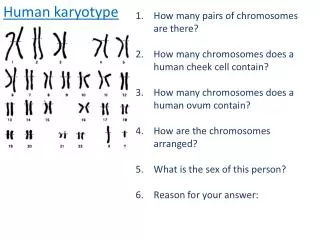
There are 2 chromosomes in each pair. • Where do the 2 members of each pair come from? • 23 from MOM • 23 from DAD Homologous Pairs
Chromosome number is constant from species to species. Humans, for example, have 46 chromosomes (23 pairs) in each body cell. Any change in chromosome number will cause a change in the amounts of proteins produced in a cell. These changes can have a considerable, detrimental effect on the individual.
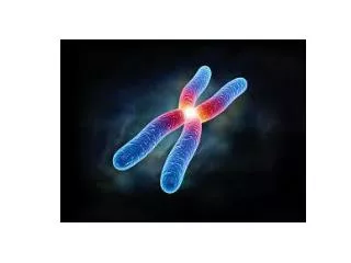
The centromere is a region of DNA where the two identical copies of DNA, formed during replication, are attached. • Each strand of replicated DNA is called a chromatid. • The chromatids on a single chromosome are called sister chromatids.
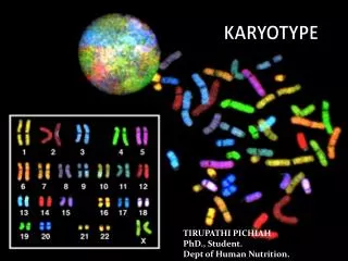
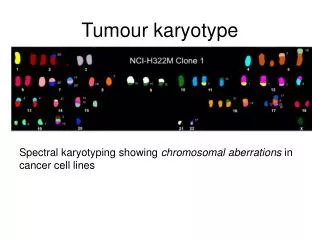
Staining the chromosomes with a dye produces the banding patterns on the chromatids.
In order to study the chromosomes of a patient, a sample of cells must first be obtained from them. Chromosome analysis can be performed using mitotic (dividing) cells from a number of sources, including white blood cells or skin cells. • Then, chemicals are added to stop the cells at a particular point in the process of cell reproduction when the double armed chromosomes can be easily seen, counted and organized into numbered groups. • The scientist then attempts to find all the homologous pairs and organize them into a picture called a karyotype. • Observing a karyotype can help us understand the nature of several genetic disorders. One of the most common times for a karyotype to be performed is during pregnancy.
Computer-assisted karyotype preparation is now commercially available. In this system a television camera and a computer are coupled to a microscope. • As chromosomes in metaphase are located, the television camera records the microscopic image, and the image is transmitted to the computer, where it can be analyzed and processed into a karyotype. • Either way, the scientist can look at the completed picture to see whether any of the chromosomes are missing or damaged or if there are any extra chromosomes.
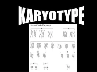
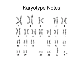
Picture representation of all of the chromosomes in a cell organized into their homologous pairs. • Reveals genetic information for that cell. What is a karyotype?
Notice that there are 22 pairs of homologous chromosomes. These are called autosomes. • The 23rd pair includes the sex chromosomes. • If a Karyotype includes two X chromosomes (XX), the individual is a female. • Males carry an X and Y chromosome (XY). Any individual with a Y chromosome is considered a male.
Amniocentesis • Chorionic Villi Sampling USING A KARYOTYPE TO DIAGNOSE CHROMOSOMAL ABNORMALITIES
16 – 20 weeks in pregnancy • Sample of cells taken from amniotic fluid • Karyotype constructed to check for abnormalities • Weeks to make karyotype • Easily detects spinal chord injuries Amniocentesis
10-12 weeks • Sample of cells taken from chorion • Hours to make karyotype • Can not detect spinal chord injuries Villi Sampling
Karyotypes constructed from cells in metaphase • If there is a problem, genetic counselors work with parents to make decisions for the best welfare of the baby Testing
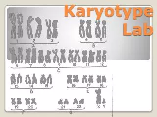
You will be given a sample chromosome spread. It will have a letter (A-F) printed at the top of the page. • Record the letter of your karyotype on the page titled “Sample Karyotype.” • Cut out each chromosome. It doesn’t have to be exact; a rectangular cut is best. • Use the normal karyotype found in this lab as a guide to identify the number of each cutout chromosome. • Carefully glue the homologous pairs of chromosomes next to each other, above the correct number of the chromosomes on the “Sample Karyotype” page. • Use the information in the next section to determine the genetic syndrome that your sample karyotype indicates. PREPARATION OF A SAMPLE KARYOTYPE
http://www.ncbi.nlm.nih.gov/pubmedhealth/PMH0002560/ Cri – du - Chat #5 upper arm deletion
http://www.ncbi.nlm.nih.gov/pubmedhealth/PMH0001992/ Down Syndrome Trisomy 21
http://www.ncbi.nlm.nih.gov/pubmedhealth/PMH0002626/ Edwards Syndrome Trisomy 18
Jacobs Syndrome XYY
http://www.ncbi.nlm.nih.gov/pubmedhealth/PMH0001420/ Kleinfelter Syndrome XXY
http://www.ncbi.nlm.nih.gov/pubmedhealth/PMH0001417/ Turner Syndrome XO