Myoglobin & Hemoglobin
Myoglobin & Hemoglobin. Structure, Function & malfunction of Biomolecules. Primary Structure. Sequence of amino acids in a protein connected via peptide linkage. Example –the enzyme lysozyme: 1 2 3 4 5 126 127 128 129 Lys-Val-Phe-Gly-Arg...Gly-Cys-Arg-Leu

Myoglobin & Hemoglobin
E N D
Presentation Transcript
Myoglobin & Hemoglobin Structure, Function & malfunctionof Biomolecules
Primary Structure Sequence of amino acids in a protein connected via peptide linkage. Example –the enzyme lysozyme: 1 2 3 4 5 126 127 128 129 Lys-Val-Phe-Gly-Arg...Gly-Cys-Arg-Leu Note: By convention, amino acid sequences are written starting with the amino terminus.
Secondary Structure Regular patterns of relatively small segments of a protein held together mainly by H-bonds Examples: -structure α-helix http://www.ultranet.com/~jkimball/BiologyPages/S/SecondaryStructure.html
Tertiary Structure Overall 3-D shape of a protein. Two basic types are globular and fibrous. Examples: Fibrous (Collagen) Globular (Pepsin)
Quaternary Structure Overall 3-D shape of a multi-subunit protein Example: Rabbit muscle glycogen phosphorylase http://bmbiris.bmb.uga.edu/wampler/tutorial/prot4.html
Protein Function in Cell • Enzymes • Catalyze biological reactions • Structural role • Cell wall • Cell membrane • Cytoplasm
Protein: The Machinery of Life NH2-Val-His-Leu-Thr-Pro-Glu-Glu- Lys-Ser-Ala-Val-Thr-Ala-Leu-Trp- Gly-Lys-Val-Asn-Val-Asp-Glu-Val- Gly-Gly-Glu-…..
Oxygen Transport Proteins • Myoglobin • Exhibits Michaelis-Menten properties • Hemoglobin • Exhibits allosteric properties
Myoglobin • Single polypeptide with 154(human) amino acids • C774H1224N210O222S5 17,183.8 daltons(human) • 8 a helices (A-H) • Located in skeletal & cardiac muscle • [high] in diving mammals like whale & seals
100 saturation with O2 50 arterial pressure tissues 2.8 20 100 pO2 (partial pressure of O2) (Torr) O2 Binding Curve • Myoglobin has high affinity for O2. • P50 = 2.8 Torr • Allows myoglobin to act as O2 storage reserve. • Releases O2 when pO2 becomes low indicating O2 deprivation.
Heme Prosthetic Group • Heme (Fe2+) has affinity for O2. • Hematin (Fe3+) cannot bind O2. • Located in crevice where it is protected from oxidation.
Oxygen Binding to Myoglobin • O2 binds to only available coordination site on iron atom. • His 93 (proximal his) binds directly to iron. • His 64 (distal his) stabilizes the O2 binding site. distal histidine proximal histidine http://cwx.prenhall.com/horton/medialib/media_portfolio/text_images/FG04_44.JPG
O O O C Fe Fe Myoblobin:CO complexes • CO binds tightly; linear. • O2 binds less tightly, bent structure. • Distal His forces bent binding of both, weakens CO binding.
Hemoglobin – Quaternary Structure • a2b2 Two α (141 AA/ α)subunits and two β (146 AA/ β)subunits
Hemoglobin Structure • Each polypeptide chain resembles myoglobin tertiary structure but 1˚ sequence varies. • Invariant residues indicate importance of those residues in function.
Oxygen Binding • Hb exhibits + cooperativity. Eaton et al. Nature Struct. Biol. 1999, 6, 351
O2 Binding to Hemoglobin Water bound to heme Iron
O2 Binding to Hemoglobin Oxygen bound to heme Iron
Hb T-state deoxy
Hb R-state - oxy
Hb Variants • HbA2 • a2d2 • Present in ~2% of adults • Embryonic Hb • a2e2 • Has affinity for O2 • Fetal Hb • a2g2 • Has affinity for O2 http://oregonstate.edu/instruction/bb450/stryer/ch10/Slide27.jpg
Bohr Effect • CO2 pH • Some side groups remain protonated at lower pH. • Stabilizes T state and promotes unloading of O2 to active tissues. • Binding of CO2 also stabilizes T state. • CO2 binds to a amino groups. http://cwx.prenhall.com/horton/medialib/media_portfolio/text_images/FG04_50.JPG
2, 3-Bisphosphoglycerate • Stabilizes deoxyHb (T state) • Facilitates unloading of O2 in tissue. 100 - BPG saturation with O2 50 + BPG 20 100 pO2 (partial pressure of O2) (Torr)
2,3-BPG Binding to Hb http://oregonstate.edu/instruction/bb450/stryer/ch10/Slide26.jpg
High Altitude and BPG • At higher altitudes, the [BPG] increases allowing Hb to unload O2 more easily. http://www.bio.davidson.edu/Courses/anphys/1999/Yusi/dpgoxyhbgraph.jpg
Stored Blood & BPG • 2,3-BPG becomes depleted in stored blood, so R state of Hb is stabilized. • If BPG depleted blood is used for a transfusion, the R state Hb doesn’t release O2. • Add inosine to stored blood to maintain BPG levels.
CO Poisoining • CO is “competitive inhibitor” of O2. • Affinity is 200X greater than that of O2. • CO also inhibits unloading O2 of in tissues.
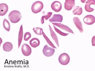
Sickle Cell Anemia Normal red blood cells are round like doughnuts, and they move through small blood tubes in the body to deliver oxygen. Sickle red blood cells become hard, sticky and shaped like sickles. When these hard and pointed red cells go through the small blood tube, they clog the flow and break apart. This can anemia.
The origin of the disease is a small change in the protein hemoglobin The change in cell structure arises from a change in the structure of hemoglobin. A single change in an amino acid causes hemoglobin to aggregate. a b
Scanning electron microscopic image of Red bllod cells
Sickle Cell Hemoglobin Significant change in structure caused by the single mutation Hemoglobin S
L-Glutamic acid(Glu/ E) L-Valine(Val / V)
Sickle Cell Hemoglobin at6β Normal mRNA GUG CAC CUG ACU CCU GAG GAG AAG val his leu thr pro glu glu lys 1 2 3 4 5 6 7 8 Normal protein Mutation (in DNA) Mutant mRNA GUG CAC CUG ACU CCU GUG GAG AAG val his leu thr pro val glu lys 1 2 3 4 5 6 7 8 Mutant protein Glutamate (glu), a negatively charged amino acid, is replaced by valine (val), which has no charge.