Download
1 / 66
880 likes | 1.66k Vues
The nervous system links sensory receptors & motor effectors in all vertebrates (and most invertebrates). Central Nervous System (CNS). Peripheral Nervous System (CNS). 23.1 Evolution of the Animal Nervous System.

E N D
The nervous system links sensory receptors & motor effectors in all vertebrates (and most invertebrates) Central Nervous System (CNS) Peripheral Nervous System (CNS) 23.1 Evolution of theAnimal Nervous System • Association neurons (or interneurons) are located in the brain and spinal cord • Motor (or efferent) neurons carry impulses away from CNS • Sensory (or afferent) neurons carry impulses to CNS
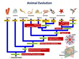
Fig. 23.3 Invertebrate Nervous Systems • Sponges are the only major phylum of multicellular animals that lack nerves • Cnidarians have simplest nervous system • Neurons are linked to one another through a nerve net • No associative activity • Just reflexes • First associative activity is seen in free-living flatworms • Two nerve cords run down bodies • Permit complex control of muscles
Fig. 23.3 • 1. More sophisticated sensory mechanisms • 2. Differentiation into central and peripheral nervous systems • Evolutionary path to vertebrates • 3. Differentiation of sensory and motor nerves • 4. Increased complexity of association • 5. Elaboration of the brain
All neurons have the same basic structure Cell body – Enlarged part containing the nucleus Dendrites – Short, slender input channels extending from end of cell body Axon – A single, long output channel extending from other end of cell body 23.2 Neurons GenerateNerve Impulses
Most neurons require nutritional support provided by companion neuroglial cells • Schwann cells (PNS) and oligodendrocytes (CNS) envelop the axon with fatty material called myelin • Myelin acts as a electrical insulator • During development cells wrap themselves around each axon several times to form a myelin sheath • Uninsulated gaps are called nodes of Ranvier • Nerve impulses jump from node to node • Multiple sclerosis and Tay-Sachs disease result from degeneration of the myelin sheath
Fig. 23.4 Structure of a neuron and formation of the myelin sheath
The Nerve Impulse • When a neuron is “at rest”, active transport channels in cell membranes move Na+ out and K+ into cells • Concentration of Na+ builds up outside the cell • K+ may diffuse out through open channels • Thus, neuron’s outside is more positive than inside • Cell membrane is said to be “polarized” • Resting potential is the charge separation between cell’s interior and exterior • – 70 millivolts
A nerve impulse results from ion movements of in and out of voltage-gated channels • A sensory input causes Na+ channels to open • Sudden influx of Na+ into cell causes “depolarization” • Local voltage change opens adjacent Na+ channels • Thus, an action potential is produced • After a slight delay, K+ voltage-gated channels open • K+ flows out of the cell • The negative charge in the cell is restored • Na+ channels snap close again • The resting potential is restored by the action of the sodium-potassium pump
Fig. 23.6 23.3 The Synapse • A synapse is the junction of an axon and another cell • Presynaptic membrane • Located on the near (axon) side of the synapse • Postsynaptic membrane • Located on the far (receiving) side of the synapse
Fig. 23.7 • Neurotransmitters are chemical messengers that carry nerve impulses across synapses • Bind to receptors in the postsynaptic cell • Cause chemically-gated channels to open
Kinds of Synapses • Excitatory synapse • Receptor protein is a chemically-gated sodium channel • On binding the neurotransmitter, the channel opens • Na+ floods inwards • Action potential begins • Inhibitory synapse • Receptor protein is a chemically-gated potassium or chloride channel • On binding the neurotransmitter, the channel opens • K+ floods outwards or Cl– floods inwards • Action potential is inhibited
Fig. 23.8a Kinds of Synapses • An individual nerve cell can possess both kinds of synapses • Integration • Various excitatory and inhibitory electrical effects cancel or reinforce one another • Occurs at the axon hillock
Neurotransmitters and Their Functions • Acetylcholine • Released at the neuromuscular junction • Have an excitatory effect on skeletal muscle and inhibitory effect on cardiac muscle • Glycine and GABA • Inhibitory neurotransmitters • Important for neural control of brain function • Biogenic amines • Dopamine – Control of body movements • Serotonin – Sleep regulation and mood
Fig. 23.9 23.4 Addictive Drugs Acton Chemical Synapses • Neuromodulators are chemicals that prolong the effect of neurotransmitters • Aid their release • Prevent their reabsorption • Example: • Depression may be caused by a shortage of serotonin • Prozac, inhibits its reabsorption
Drug Addiction • A cell that is exposed to a chemical signal for a prolonged time, loses its “sensitivity” • It tends to lose its ability to respond to the stimulus with its original intensity • Nerve cells are particularly prone to this loss of sensitivity • They respond to high neurotransmitter exposure by inserting fewer receptor proteins
Drug Addiction • Cocaine is a neuromodulator • It causes large amounts of neurotransmitter to remain in synapses for long periods of time • Dopamine transmits pleasure messages in the body’s limbic system • High levels for long periods of time, cause nerve cells to lower the number of receptors • Addiction occurs when chronic exposure to a drug induces the nervous system to act physiologically
Addiction to Smoking • “Nicotine receptors” normally served to bind acetylcholine • Brain adjusts to prolonged exposure to nicotine by • 1. Making fewer nicotine receptors • 2. Altering the pattern of activation of nicotine receptors • Addiction occurs because the brain compensates for the nicotine-induced changes by making others • There is no easy way out • The only way to quit is to quit!
Fig. 23.12 23.5 Evolution of the Vertebrate Brain • Brains of primitive fish, while small, already had the 3 divisions found in contemporary vertebrate brains • 1. Hindbrain • Rhombencephlon • 2. Midbrain • Mesencephlon • 3. Forebrain • Prosencephlon
Hindbrain • Major component of early fishes, as it is today • An extension of the spinal cord devoted primarily to coordinating muscle reflexes • Most coordination is done by the cerebellum • Midbrain • Composed primarily of optic lobes • Receive and process visual information • Forebrain • Devoted for processing olfactory (smell) information • Note: • Brains of fishes continue growing throughout their lives!
Diencephalon • Thalamus – Relay center between incoming sensory information and the cerebrum • Hypothalamus – Coordinates nervous and hormonal responses to many internal stimuli and emotions • Telencephalon • Devoted largely to associative activity • Cerebrum (mammals) • Dominant part of the brain • Receives sensory data and issues motor commands • Starting with the amphibians, sensory information is increasingly centered in the forebrain
Fig. 23.13 The evolution of the vertebrate brain Cerebrum dominance is greatest
Fig. 23.14 23.6 How the Brain Works • Cerebrum is ~ 85% of the weight of the human brain • Functions in language, thought, personality and other “thinking and feeling” activities • Much of activity occurs in the cerebral cortex • Gray outer layer
The cerebrum is divided by a groove into right and left halves called cerebral hemispheres • Linked by bundles of neurons calledtracts • Serve as information highways • In general, the left brain is associated with language, speech and mathematical abilities • The right brain is associated with intuitive, musical, and artistic abilities • Stroke • A disorder caused by blood clots blocking blood vessels in the brain
Fig. 23.16 • Thalamus • Major site of sensory processing in the brain • Controls balance • Hypothalamus • Integrates internal activities • Body temperature, blood pressure, etc. • Controls pituitary gland secretions • Linked to areas of cerebral cortex via limbic system Center for pain, anger, sex, hunger, etc. Memory center • Responsible for deep-seated drives and emotions
Cerebellum • Extends back from the base of the brain • Coordinates muscle movement • Even better developed in birds • Brain Stem • Made up of midbrain, pons, and medulla oblongata • Connects rest of brain to spinal cord • Controls breathing, swallowing, digestion • As well as heart beat and blood vessel diameter
Language and other higher functions • Left hemisphere is “dominant” hemisphere for language • It is adept at sequential reasoning • The “nondominant” hemisphere (the right hemisphere in most people) is involved in • Spatial reasoning (assembling puzzles) • Musical ability • Short-term memory appears to be stored electrically in the form of a transient neural excitation • Long-term memory appears to involve structural changes in certain neural connections
Alzheimer Disease • Memory and thought processes become dysfunctional • Two hypotheses have been proposed for the cause • 1. Brain nerve cells are killed from the outside in • Accumulation of plaques of abnormal external proteins called b-amyloid peptides • 2. Brain nerve cells are killed from the inside out • Accumulation of tangles of abnormal internal proteins called tau (t) • Researchers continue to study whether tangles and plaques are causes or effects of Alzheimer disease
23.7 The Spinal Cord • The spinal cord is a cable of neurons extending from the brain down through the backbone • Neuron cell bodies in the center • Gray matter • Axons and dendrites on the outside • White matter • It is surrounded and protected by the vertebrae • Through them spinal nerves pass out to the body • Motor nerves from spine control most of the muscles below the head
Fig. 23.20 23.8 Voluntary and AutonomicNervous Systems Are two subdivisions of vertebrate motor pathways
Fig. 23.21 The knee-jerk reflex The voluntary nervous system relays commands to skeletal muscles • Can be controlled by conscious thought Reflexes are sudden involuntary movements • Are rapid because sensory neuron passes information directly to a motor neuron • Most involve single connecting interneuron between sensory and motor neurons
The autonomic nervous system stimulates glands andrelays commands to smooth muscles • Cannot be controlled by conscious thought • Composed of elements that act in opposition to each other • Sympathetic nervous system • Dominates in time of stress • Controls the “fight-or-flight” reaction • Increases blood pressure, heart rate, breathing • Parasympathetic system • Conserves energy by slowing down processes
Fig. 23.22 How the sympathetic and parasympathetic nervous systems interact
Fig. 23.23 Kangaroo rats have specialized ears 23.9 Sensory Perception • The sensory nervous system tells the central nervous system what’s happenin’! Adapted to nocturnal life • Sensory receptors • Specialized sensory cells that detect changes inside and outside the body • Sensory organs • Complex sensory receptors • Eyes, ears, taste buds
The path of sensory information is a simple one • 1. Stimulation • Physical stimulus impinges on a sensory receptor • 2. Transduction • Stimulus-gated ion channels in sensory neuron are opened or closed • An action potential is generated • 3. Transmission • Nerve impulse is conducted to the CNS • Two main types of sensory receptors • Extroreceptors sense stimuli in external environment • Introreceptors sense stimuli in internal environment
Sensing the Internal Environment • Vertebrates use many different sensory receptors to respond to changes in internal environment • Temperature Change • Two nerve endings in the skin • One stimulated by cold, the other by warmth • Blood chemistry • Receptors in arteries sense blood CO2 levels • Pain • Special nerve endings within tissues near the surface
Fig. 23.24 Fig. 23.25 • Muscle contraction • Sensory receptors embedded within muscle • Touch • Pressure receptors buried below skin • Blood pressure • Neurons called baroreceptors in major arteries
23.10 Sensing Gravity and Motion • Receptors in the ear inform the brain where the body is in three dimensions • Balance • Gravity is detected byshifting of otolith sensory receptors • These are located in a gelatin-like matrix in the utricle and saccule chambers of the inner ear • Motion • Motion is detected by the deflection of hair cells by fluid in a direction opposite to that of motion • These hair cells are found in the cupula • Tent-like assemblies in the three semicircular canals
Fig. 23.27 23.11 Sensing Chemicals:Taste and Smell • Taste • Taste buds arelocated in raised areas called papillae • Food chemicals dissolve in saliva and contact the taste cells
Fig. 23.28 23.11 Sensing Chemicals:Taste and Smell • Smell • Olfactory receptor cellsare embedded in the epithelium of the nasal passage • These are far more sensitive in dogs than in humans
23.12 Sensing Sounds: Hearing • When a sound is heard, air vibration is detected • Eardrum membrane is pushed in and out by waves of air pressure • Three small bones (ossicles) located on other side of eardrum increase the vibration force • Amplified vibration is transferred to fluid within the inner ear • Inner ear chamber is shaped like a tightly coiled snail shell and is called cochlea
Cochlea sound receptors are hair cells that rest on a membrane running up and down the chamber • They are covered by another membrane • Sound waves entering the cochlea cause this membrane “sandwich” to vibrate • Bent hair cells send nerve impulses to brain • Sounds of different frequencies cause different parts of the membrane to vibrate • Different sensory neurons are fired • Sound intensity is determined by how often the neurons fire