Download
1 / 35
350 likes | 390 Vues
Delve into the intricate world of developmental neurobiology to comprehend the construction, function, and repair of the nervous system. This scientific field examines neuronal diseases, plasticity, and evolution to reveal how neurons develop, wire, and adapt within the brain. Covering neuronal origins, identity, specificity, and plasticity, this study reveals the remarkable journey of nervous system development. Explore key stories, mechanisms, and model organisms in neurodevelopment with a focus on Genomics impact. Join us on a captivating journey through the embryology of the nervous system, from germ bands to cell differentiation, apoptotic processes, and neuronal connections patterning. Through detailed lectures, discussions on growth cones, axonal guidance cues, and in-depth exploration of vertebrate embryology, discover the fascinating world of developmental neurobiology.

E N D
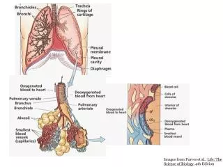
Developmental Neurobiology • Textbook Readings: • (“Neuroscience, 3rd Edition”, Purves, et al.) • Chapter 1 Studying Nervous Systems • 7 Intracellular Signal Transduction • 21 Early Brain Development • 22 Construction of Neural Circuits • (23) Modification of Developing Brain • Circuits by Neural Activity • (24) Plasticity in the Adult Nervous • System • References: • “Principles of Neural Development”, • Dale Purves and Jeff Lichtman, Sinauer Press. • 2) “Developmental Biology, 5th Edition”, • Scott Gilbert, Sinauer Press.
Why Study Developmental Neurobiology? • Terrific scientific challenge. • To understand the construction and normal • operation of the developing (and adult) • nervous system. • To understand human neuronal diseases • and the repair and regeneration of the • nervous system. • To understand neuronal plasticity / • learning and memory. • To understand the evolution of nervous • systems.
Emphases of Lectures • Neurodevelopment survey: • Highlights of a few key stories. • Cellular and molecular mechanisms. • Synergy of observational and • experimental science. • Model organisms. • Impact of Genomics.
Drosophila Optic Lobe (Courtesy of N. Strausfeld, U. Arizona)
Neuronal Circuits Contain Diverse Neurons Which Must Be Wired Exactly. Spatiotopy Stereotopy VS1 (Borst and Egelhaaf, 1992; Scott, Raabe, Luo, 2002) Me Me Lo Lo La La
Embryonically Regulated Genes are the Most Abundant Genes in the Drosophila Genome (Most of these are Neuronal Genes) (Abeitman, et al., 2002)
Major Questions 1. Origins. Where do neurons come from? 2. Identity. How does a neuron know what to be? 3. Specificity. How does a neuron make the right connections? 4. Plasticity. How does the nervous system adapt to mistakes and experience?
Development proceeds by progressive developmental restrictions. (pluripotent, stem cell) (differentiated)
Developmental Restrictions may be: 1) Genetic (programmed by genes) or 2) Epigenetic (determined by environment). (pluripotent, stem cell) (genetic) (environmental) (differentiated)
Recipe for Constructing a Nervous System Selective Assortment Proliferation Morphogenesis (& Segmentation) Specification of Identity Modification by Cell Death & Experience (Plasticity) Axonal Outgrowth & Synapse Formation
Specific Topics • Lecture 17 (Mon Oct 30) • Embryology of the Nervous System • Germ bands and cell types. • Body axes. • Folding, involutions and morphogenic movements. • Origin, migration and differentiation of neurons. • Lecture 18 (Wed Nov 3) • Embryonic Neural Induction • Spemann organizer. • Molecular mechanisms of induction (TGF-b). • Intra- and intermolecular signaling. • Lecture 19 (Mon Nov 6, Wed Nov 8) • Cell Death (Apoptosis) • Cell death and trophic factors (NGF) in developing NS. • Molecular mechanism of apoptosis. • Lecture 20 (Mon Nov 13) • Patterning Neuronal Connections • Growth Cones. • Axonal guidance cues.
Vertebrate Embryology (Frog) 1. The unfertilized oocyte contains positional information (cytoplasmic determinants) contributed maternally. Some maternal RNAs are not equally distributed. The first informational axis is intrinsic to oocyte! Animal Vegetal (yolk)
2. Fertilization triggers an influx of calcium, that sweeps across the egg. This causes rapid release of cortical granules, forming the fertilization envelope, blocking polyspermy. Animal Cortical granules Sperm entry point Ca+2 Vegetal (yolk)
3. The point of entry of the sperm creates a second positional axis, dorsal (opposite side of entry point) and ventral (entry point). Animal Sperm entry point Cortical Rotation mixes cytoplasmic determinants and creates the dorsal-ventral axis. Vegetal (yolk) Animal Gray Crescent (future blastopore) Ventral Dorsal future Nieuwkoop Center Vegetal (yolk)
Time-Lapse Videos Gray Crescent Formation in Xenopus (Courtesy of Jeffrey Hardin, University of Wisconsin)
Cortical Rotation Leads to Unequal Combinations of Cytosolic Maternal Determinants which Partition into Dividing Embryonic Cells Gray Crescent Nieuwkoop Center
Early Cell Divisions of an Amphibian Embryo to Create the Blastula. blastomere blastocoel Blastula
(Animal) (Ventral) future Blastopore Site of sperm entry (Dorsal) (Vegetal) Fate Mapping the Blastula: 3 Major Spatial Axes Formed by Gradients of Signaling Molecules. 1. Animal/Vegetal (Maternal Determinants) 2. Dorsal/Ventral (Sperm Entry, Cortical Rotation)
(Anterior) (Posterior) Spemann Organizer Blastopore Nieuwkoop Center Fate Mapping the Blastula: 3 Major Spatial Axes Formed by Gradients of Signaling Molecules; Nieuwkoop Center Induces the Spemann Organizer. 1. Animal/Vegetal (Maternal Determinants) 2. Dorsal/Ventral (Sperm entry, Cortical Rotation) 3. Anterior/Posterior(Spemann Organizer)
(Anterior) Ectoderm (Skin, Neurons) (Animal) (Posterior) (Ventral) Blastopore (Dorsal) Mesoderm (Notocord, Muscle, Bone, Blood) (Vegetal) Endoderm (Lining of Gut, Placenta in Mammals) Fate Map of the Blastula: 3 Principle Germ Bands Created.
Gastrulation of the Amphibian Blastula.
Gastrulation of Xenopus Blastula: 3-D Microscopic (Confocal) Reconstruction. (Ewald, et al., 2004)
Neural plate (Apposition of Different Germbands) Blastopore and yolk plug • Endoderm and Mesoderm • Involute with Gastrulation. • Mesoderm Apposes • Overlying (Neuro)Ectoderm, • and Induces the Neural Plate.
Gastrulation and Neurulation in Xenopus Early Cell Divisions in Zebrafish embryo (Courtesy of Jeffrey Hardin, University of Wisconsin) (Courtesy of Paul Myers, University of Minnesota) Time-Lapse Videos
Ant Post D V Formation of Neural Crest Cells (makes PNS, endocrine cells, pigment cells, connective tissue). Closure of the neural tube. Neurulation.
Neural Crest Neurulation: Origin of Floor Plate and Neural Crest.
The earliest born neurons are found closest to the ventricular surface (thymidine pulse-chase labeling of dividing cells). Neurons are born in the ventricular layer and migrate radially along glia to their differentiated adult cortical layer. (Rakic, 1974) Cortical Development: Laminar structure of the cortex is constructed from the inside-out.
Isolecithal eggs • (protochordates, mammals): 2. Mesolecithal eggs (amphibians): 3. Telolecithal eggs (reptiles, birds, fish): (epiboly) (blastodisc) The amount of yolk determines the symmetry of early cleavages and the shape of the blastula.
4. Mammalian eggs have no yolk, so early divisions resemble isolecithal eggs (protochordate-like). However, later stages resemble the blastodisc of telolecithal eggs (reptile/bird/fish-like). a) Blastula flattens into the inner cell mass. b) Endodermal cells form the trophoblast and placental structures.
Key Points of Lecture 17: 1. Three germbands, ectoderm (skin and neurons), mesoderm (muscle, blood and internal organs) and endoderm (lining of the gut). 2. Development proceeds from pleuripotency (stem cells) to the differentiated state (adult neuron). 3. Neuronal induction requires specific contact between groups of cells; embryonic morphogenesis allows this occur. 4. Positional information is created early by asymmetric distribution of molecules. These form axes (Animal/Veg, D/V, Ant/Post) that guide the movement of embryonic cells.
Key Points of Lecture 17 (cont): 5. Key morphological landmarks of embryogensis: a) Fertilization/Cortical Rotation. b) Blastula (hollow ball of cells). c) Gastrulation (inside-out involution of surface cells to the interior, through the blastopore). d) Neurulation (neural tube formation). e) Segmentation/Cephalization. f) Birth of neurons from the ventricular zone. Radial, then tangential migration to final destination.
Embryogenesis (Key Steps): 1. Oocyte possess maternal cytoplasmic determinants. 2. Fertilization triggers calcium influx, and creates dorsal-ventral axis by cortical rotation. 3. Cell divisions, synchronous at first, then asynchronous. 4. Blastula created. (“Hollow” ball of cells) 6. Germ bands (ectoderm, mesoderm, endoderm) created by molecular signals along the Animal/Vegetal axis. 5. Gastulation. (Involution of superficial cells through the blastopore). 6. Anterior-posterior axis created by Spemann organizer.
Embryogenesis (Key Steps) (cont.): 7. Apposition of future mesoderm with neuroectoderm induces the neural plate. 8. Neurulation. (Lateral neural folds bend over the midline and fuse into the neural tube.) 9. Neural crest cells derived from leading edge of neural folds, migrate into somites to form the PNS. 10. Segmentation, anterior enlargement (cephalization), cortical development and spinal specification.