Download
1 / 21
370 likes | 1.39k Vues
INSPECTION AND PALPATION OF THE PRECORDIUM. Hakan Karpuz, MD Dept. of Cardiology Cerrahpaşa Medical School. Physical Examination. 1- Inspection 2- Palpation 3- Percussion 4- Oscultation. ... first evaluation of the patients for diagnosis of

E N D
INSPECTION AND PALPATION OF THE PRECORDIUM Hakan Karpuz, MD Dept. of Cardiology Cerrahpaşa Medical School
PhysicalExamination 1-Inspection 2-Palpation 3-Percussion 4-Oscultation
... first evaluation of the patients for diagnosis of cardiovascular disease begins with the first visual approach ...
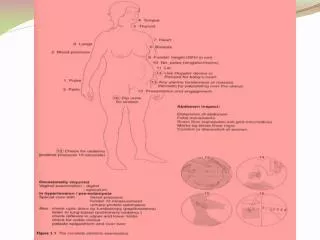
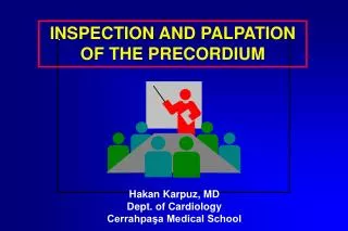
Precordial inspection The cardiac examination proper should commence with inspection of the chest, which can best be accomplished with the examiner standing at the foot of the bed or examining table.
Precordial inspection build obesity, Marfanoid, heavy muscular thorax (contrasting with lessdevelopped lower extremities)- respiration: frequency, regularity and depth - cutaneous abnormality: dilation of veins,
Precordial inspection scars: sternotomy, valvotomy, … - deformity: pectus excavatum, pectus carinatum (pigeon chest) - pulsations: “danse des arteres”
Precordial inspection blue sclera aortic dilatation- conjuctival bleedinginfective endocarditis- arcus senilishiperlipidemia
Precordial inspection clubbing CHD, pulmonary hypertension- leg oedema congestive heart failure
Precordial palpation This checks in:- left parasternal area- right parasternal area - cardiac apex
Precordial palpation Left parasternal area :- 2. left intercostal space: pulmonary artery - 3.-5. left intercostal space: right ventricle’s activity left atrium some murmurs
Precordial palpation Right parasternal area :- 1. right intercostal space: aorta- 2. right intercostal space: aortic valve, hypertension - 3.-5. right intercostal space: right atrium
Precordial palpation Cardiac apex :- 5. left intercostal space (left midclavicular line): left ventricular contraction
Precordial palpation This checks for:- thrills- apex beat - palpable sounds - abnormal pulsation
Precordial palpation (thrills) Systolic thrill- aortic area: aortic stenosis- left sternal edge: ventricular septal defect- apex: ruptured mitral chordea- pulmonary area: pulmonary stenosis- subclavicular area: subclavian stenosis
Precordial palpation (thrills) Diastolic thrill(less common)- apex: mitral stenosis (patient lying on left side and breath held in expiration)- left sternal edge: aortic regurgitation (occasionally)
Precordial palpation (apex beat) Cardiac apical impulse is normally localized in the fifth left intercostal space, midclavicular line; It is palpable but does not lift the finger off the chest.Abnormalities- forceful apical thrust: left ventricular hypertrophy- lateral and downward displacement of apex impulse: left ventricular dilatation
Precordial palpation (apex beat) - prominent presystolic impulse: hypertension, aortic stenosis - double systolic apical impulse: hypertrophic cardiomyopathy- sustained “lift” at lower left sternal border right ventricular hypertrophy- dyskinetic (outward bulge) impulse: ventricular aneurysme, cardiomyopathy
Precordial palpation (palpable sounds) Palpable heart sounds represent forceful valve closure, or valve situated close to the chest wall, e.g. palpable- S1 (mitral closure) in mitral stenosis- P2 in pulmonary hypertension- A2 in transposition- both S1 and S2 in thin patients with tachycardia
Precordial palpation (abnormal pulsation) Abnormal pulsations are very variable, e.g.- ascending aortic aneurysm pulsating in aortic area- right ventricular outflow tract aneurysm in pulmonary area- collateral pulsation round the back in coarctation- pulsatile right ventricular outflow tract in atrial septal defect
Precordial percussion Percussion of cardiac dullness is not clinically very useful; the Rx gives a better idea of heart size.
Tıp teknolojisinin iyi bir fizik muayene ile kombinasyonu, tanı yetersizlikleri ve aşırı tetkik isteme alışkanlığından özellikle genç hekimleri kurtaracaktır.